YAP1 Polyclonal antibody 13584-1-AP proteintech
$149.00
In stock
SKU
13584-1-AP
| Catalog Number | 13584-1-AP |
| Synonyms | 65 kDa Yes associated protein, Protein yorkie homolog, Transcriptional coactivator YAP1, YAP, yes associated protein 1 |
| Host | Rabbit |
| Reactivity | human, mouse, rat and More (5) |
| Reactivity | human, mouse, rat, pig, rabbit, monkey, chicken, zebrafish |
| Form | Liquid |
| Formulation | PBS, Azide, Glycerol |
| Applications | WB, IHC, IF/ICC, IF-P, IP, CoIP, ChIP, ELISA |
| Applications | WB, IHC, IF/ICC, IF-P, IP, ELISA |
| Host / Isotype | Rabbit / IgG |
| Tested Applications — Positive WB detected in | HEK-293T cells, MCF-7 cells, HeLa cells, BGC-823 cells, HepG2 cells, SGC-7901 cells, mouse liver tissue, rat liver tissue, L02 cells |
| Tested Applications — Positive IP detected in | NIH/3T3 cells |
| Tested Applications — Positive IHC detected in | human liver cancer tissue, human ovary tumor tissue, human colon cancer tissue Note: suggested antigen retrieval with TE buffer pH 9.0; (*) Alternatively, antigen retrieval may be performed with citrate buffer pH 6.0 |
| Tested Applications — Positive IF-P detected in | human lung cancer tissue |
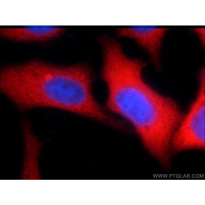
| Tested Applications — Positive IF/ICC detected in | HepG2 cells |
| Recommended Dilutions — Western Blot (WB) | WB : 1:20000-1:100000 |
| Recommended Dilutions — Immunoprecipitation (IP) | IP : 0.5-4.0 ug for 1.0-3.0 mg of total protein lysate |
| Recommended Dilutions — Immunohistochemistry (IHC) | IHC : 1:50-1:500 |
| Recommended Dilutions — Immunofluorescence (IF)-P | IF-P : 1:50-1:500 |
| Recommended Dilutions — Immunofluorescence (IF)/ICC | IF/ICC : 1:50-1:500 |
| Positive WB detected in | HEK-293T cells, MCF-7 cells, HeLa cells, BGC-823 cells, HepG2 cells, SGC-7901 cells, mouse liver tissue, rat liver tissue, L02 cells |
| Positive IP detected in | NIH/3T3 cells |
| Positive IHC detected in | human liver cancer tissue, human ovary tumor tissue, human colon cancer tissue Note: suggested antigen retrieval with TE buffer pH 9.0; (*) Alternatively, antigen retrieval may be performed with citrate buffer pH 6.0 |
| Positive IF-P detected in | human lung cancer tissue |
| Positive IF/ICC detected in | HepG2 cells |
| Western Blot (WB) | WB : 1:20000-1:100000 |
| Immunoprecipitation (IP) | IP : 0.5-4.0 ug for 1.0-3.0 mg of total protein lysate |
| Immunohistochemistry (IHC) | IHC : 1:50-1:500 |
| Immunofluorescence (IF)-P | IF-P : 1:50-1:500 |
| Immunofluorescence (IF)/ICC | IF/ICC : 1:50-1:500 |
| Tested Reactivity | human, mouse, rat |
| Cited Reactivity | human, mouse, rat, pig, rabbit, monkey, chicken, zebrafish |
| Class | Polyclonal |
| Type | Antibody |
| Immunogen | CatNo: Ag4510 Product name: Recombinant human YAP1 protein Source: e coli. -derived, PGEX-4T Tag: GST Domain: 155-504 aa of BC038235 Sequence: PTAQHLRQSSFEIPDDVPLPAGWEMAKTSSGQRYFLNHIDQTTTWQDPRKAMLSQMNVTAPTSPPVQQNMMNSASGPLPDGWEQAMTQDGEIYYINHKNKTTSWLDPRLDPRFAMNQRISQSAPVKQPPPLAPQSPQGGVMGGSNSNQQQQMRLQQLQMEKERLRLKQQELLRQAMRNINPSTANSPKCQELALRSQLPTLEQDGGTQNPVSSPGMSQELRTMTTNSSDPFLNSGTYHSRDESTDSGLSMSSYSVPRTPDDFLNSVDEMDTGDTINQSTLPSQQNRFPDYLEAIPGTNVDLGTLEGDGMNIEGEELMPSLQEALSSDILNDMESVLAATKLDKESFLTWL Predict reactive species |
| Full Name | Yes-associated protein 1, 65kDa |
| Calculated Molecular Weight | 504 aa, 54 kDa |
| Observed Molecular Weight | 65-75 kDa |
| GenBank Accession Number | BC038235 |
| Gene Symbol | YAP1 |
| Gene ID (NCBI) | 10413 |
| RRID | AB_2218915 |
| Conjugate | Unconjugated |
| Purification Method | Antigen affinity purification |
| UNIPROT ID | P46937 |
| Storage Buffer | PBS with 0.02% sodium azide and 50% glycerol, pH 7.3. |
| Storage Conditions | Store at -20°C. Stable for one year after shipment. Aliquoting is unnecessary for -20 o C storage. 20ul sizes contain 0.1% BSA. |
| IF protocol for YAP1 antibody 13584-1-AP | Download protocol |
| IHC protocol for YAP1 antibody 13584-1-AP | Download protocol |
| IP protocol for YAP1 antibody 13584-1-AP | Download protocol |
| WB protocol for YAP1 antibody 13584-1-AP | Download protocol |
| mouse | IHC Nature Divergent transcriptional regulation of astrocyte reactivity across disorders. Authors - Joshua E Burda View Article |
| human | IF Nature The SWI/SNF complex is a mechanoregulated inhibitor of YAP and TAZ. Authors - Lei Chang View Article |
| Mounika (Verified Customer) (12-25-2025) | This antibody played very critical role in our project Hippo signaling pathway, worked very excellently, enjoyed working with it at ease! Applications: Western Blot, Immunohistochemistry, Immunofluorescence, Immunoprecipitation, ChIP Primary Antibody Dilution: 1:1000 |
| Ayse (Verified Customer) (10-08-2025) | Works well. Applications: Immunohistochemistry Primary Antibody Dilution: 1:200 |
| Udesh (Verified Customer) (06-17-2025) | Worked well for IF and WB Applications: Western Blot, Immunofluorescence Primary Antibody Dilution: 1:1000 for WB and 1:2000 for IF Cell Tissue Type: MSCs |
| Kis (Verified Customer) (02-21-2025) | This antibody performs well for both Western blot (WB) and immunohistochemistry (IHC) in human and mouse models. Applications: Western Blot, Immunohistochemistry Primary Antibody Dilution: 1.1000 |
| Kazuaki (Verified Customer) (01-28-2025) | Works well Applications: Western Blot Primary Antibody Dilution: 1:4000 Cell Tissue Type: Mouse myoblast C2C12 cell line |
| Siddharth (Verified Customer) (08-04-2023) | Great for IF. Detects both Cytoplasmic and Nuclear YAP1. Applications: Immunofluorescence Primary Antibody Dilution: 1:100 Cell Tissue Type: hMSC cell line YAP1 Antibody Immunofluorescence validation (1:100 dilution) in hMSC cell line (Cat no:13584-1-AP) |
| Chiara (Verified Customer) (09-15-2022) | In RH4 cells I observe by Western blot 4 bands; two bands around 70 KDa correspond to YAP, while a single band around 55 KDa corresponds to TAZ protein as proved by a RNA interference experiment. Applications: Western Blot Primary Antibody Dilution: 1:1000 Cell Tissue Type: RH4 Rhabdomyosarcoma cell line YAP1 Antibody Western Blot validation (1:1000 dilution) in RH4 Rhabdomyosarcoma cell line (Cat no:13584-1-AP) |
| María (Verified Customer) (02-08-2022) | Works for WB (1:1000). Applications: Western Blot Primary Antibody Dilution: 1:1000 |
| Arianna (Verified Customer) (03-01-2019) | Genetically validated on YAP-null liver sections. Applications: Immunofluorescence, Primary Antibody Dilution: 1:100 Cell Tissue Type: Liver tissue |
| Joshua (Verified Customer) (12-20-2018) | Cells were fixed in 4% paraformaldehyde and stained overnight at 4C. Cells were counterstained with phalliodin and DAPI. Staining is mix of nuclear, junctional, and cytosolic. Applications: Immunofluorescence, Primary Antibody Dilution: 1:500 Cell Tissue Type: MDCK Canine Kidney Epithelial YAP1 Antibody Immunofluorescence, validation (1:500 dilution) in MDCK Canine Kidney Epithelial (Cat no:13584-1-AP) |
| Juan Pablo (Verified Customer) (11-29-2018) | CGN treated with Shh (1ug/ml) for 48hs to induced proliferation, I see a nice induction of YAP1I get a nice clean band Applications: Western Blot, Primary Antibody Dilution: 1:1000 Cell Tissue Type: Culture of Granule cell neurons from P7 rat pups YAP1 Antibody Western Blot, validation (1:1000 dilution) in Culture of Granule cell neurons from P7 rat pups (Cat no:13584-1-AP) |
| kk (Verified Customer) (11-21-2018) | Applications: Western Blot, Primary Antibody Dilution: 1:2000 Cell Tissue Type: human PANC1 cells |
| Citations | 537 |
| Dilutions | WB : 1:20000-1:100000 IP : 0.5-4.0 ug for IP and 0.5-4.0 ug for 1.0-3.0 mg of total protein lysate for WB IHC : 1:50-1:500 IF-P : 1:50-1:500 IF/ICC : 1:50-1:500 |
| Isotype | IgG |
| Clonality | Polyclonal |
| Clone Number | - |
| Conjugation | Unconjugated |
Yes-associated protein 1 (YAP1) is a transcriptional regulator which can act both as a coactivator and a corepressor and is the critical downstream regulatory target in the Hippo signaling pathway that plays a pivotal role in organ size control and tumor suppression by restricting proliferation and promoting apoptosis. The core of this pathway is composed of a kinase cascade wherein STK3/MST2 and STK4/MST1, in complex with its regulatory protein SAV1, phosphorylates and activates LATS1/2 in complex with its regulatory protein MOB1, which in turn phosphorylates and inactivates YAP1 oncoprotein and WWTR1/TAZ. Plays a key role to control cell proliferation in response to cell contact. Phosphorylation of YAP1 by LATS1/2 inhibits its translocation into the nucleus to regulate cellular genes important for cell proliferation, cell death, and cell migration. The presence of TEAD transcription factors are required for it to stimulate gene expression, cell growth, anchorage-independent growth, and epithelial mesenchymal transition (EMT) induction. Isoform 2 and isoform 3 can activate the C-terminal fragment (CTF) of ERBB4 (isoform 3).Increased expression seen in some liver and prostate cancers (PMID: 31613226, 32488048, 33520338). Isoforms lacking the transactivation domain found in striatal neurons of patients with Huntington disease (at protein level).It is actived by phosphorylation and degradated by ubiquitination (PMID: 20048001).This antibody is a rabbit polyclonal antibody. The calcualted molecular weight of YAP1 is 54 kDa, but routinely observed at 65-75 kDa by Western Blot (PMID: 28230103, 33264286, 36255405). Protocols Product Specific Protocols IF protocol for YAP1 antibody 13584-1-AP Download protocol IHC protocol for YAP1 antibody 13584-1-AP Download protocol IP protocol for YAP1 antibody 13584-1-AP Download protocol WB protocol for YAP1 antibody 13584-1-AP Download protocol Standard Protocols Click here to view our Standard Protocols
Publications