Cytochrome c Monoclonal antibody 66264-1-Ig proteintech
$149.00
In stock
SKU
66264-1-Ig
| Catalog Number | 66264-1-Ig |
| Synonyms | 2D8D11, CYC, CYCS |
| Host | Mouse |
| Reactivity | human, mouse, rat and More (2) |
| Reactivity | human, mouse, rat, pig, canine |
| Form | Liquid |
| Formulation | PBS, Azide, Glycerol |
| Applications | WB, IHC, IF/ICC, FC (Intra), ELISA |
| Host / Isotype | Mouse / IgG2a |
| Tested Applications — Positive WB detected in | HeLa cells, human heart tissue, human skeletal muscle tissue, rat skeletal muscle tissue, mouse skeletal muscle tissue, HEK-293 cells, HepG2 cells, MCF-7 cells, Jurkat cells, HSC-T6 cells, ROS1728 cells, RAW 264.7 cells |
| Tested Applications — Positive IHC detected in | human liver cancer tissue, human breast cancer tissue Note: suggested antigen retrieval with TE buffer pH 9.0; (*) Alternatively, antigen retrieval may be performed with citrate buffer pH 6.0 |
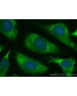
| Tested Applications — Positive IF/ICC detected in | HepG2 cells |
| Tested Applications — Positive FC (Intra) detected in | HepG2 cells |
| Recommended Dilutions — Western Blot (WB) | WB : 1:5000-1:50000 |
| Recommended Dilutions — Immunohistochemistry (IHC) | IHC : 1:1000-1:5000 |
| Recommended Dilutions — Immunofluorescence (IF)/ICC | IF/ICC : 1:200-1:800 |
| Recommended Dilutions — Flow Cytometry (FC) (INTRA) | FC (INTRA) : 0.20 ug per 10^6 cells in a 100 µl suspension |
| Positive WB detected in | HeLa cells, human heart tissue, human skeletal muscle tissue, rat skeletal muscle tissue, mouse skeletal muscle tissue, HEK-293 cells, HepG2 cells, MCF-7 cells, Jurkat cells, HSC-T6 cells, ROS1728 cells, RAW 264.7 cells |
| Positive IHC detected in | human liver cancer tissue, human breast cancer tissue Note: suggested antigen retrieval with TE buffer pH 9.0; (*) Alternatively, antigen retrieval may be performed with citrate buffer pH 6.0 |
| Positive IF/ICC detected in | HepG2 cells |
| Positive FC (Intra) detected in | HepG2 cells |
| Western Blot (WB) | WB : 1:5000-1:50000 |
| Immunohistochemistry (IHC) | IHC : 1:1000-1:5000 |
| Immunofluorescence (IF)/ICC | IF/ICC : 1:200-1:800 |
| Flow Cytometry (FC) (INTRA) | FC (INTRA) : 0.20 ug per 10^6 cells in a 100 µl suspension |
| Tested Reactivity | human, mouse, rat |
| Cited Reactivity | human, mouse, rat, pig, canine |
| Class | Monoclonal |
| Type | Antibody |
| Immunogen | CatNo: Ag24349 Product name: Recombinant human Cytochrome c protein Source: e coli. -derived, PET28a Tag: 6*His Domain: 1-105 aa of BC009578 Sequence: MGDVEKGKKIFIMKCSQCHTVEKGGKHKTGPNLHGLFGRKTGQAPGYSYTAANKNKGIIWGEDTLMEYLENPKKYIPGTKMIFVGIKKKEERADLIAYLKKATNE Predict reactive species |
| Full Name | cytochrome c, somatic |
| Calculated Molecular Weight | 12 kDa |
| Observed Molecular Weight | 12-15 kDa |
| GenBank Accession Number | BC009578 |
| Gene Symbol | Cytochrome c |
| Gene ID (NCBI) | 54205 |
| RRID | AB_2716798 |
| Conjugate | Unconjugated |
| Purification Method | Protein A purification |
| UNIPROT ID | P99999 |
| Storage Buffer | PBS with 0.02% sodium azide and 50% glycerol, pH 7.3. |
| Storage Conditions | Store at -20°C. Stable for one year after shipment. Aliquoting is unnecessary for -20 o C storage. 20ul sizes contain 0.1% BSA. |
| FC protocol for Cytochrome c antibody 66264-1-Ig | Download protocol |
| IF protocol for Cytochrome c antibody 66264-1-Ig | Download protocol |
| IHC protocol for Cytochrome c antibody 66264-1-Ig | Download protocol |
| WB protocol for Cytochrome c antibody 66264-1-Ig | Download protocol |
| mouse,human | WB Hepatology Hepatic stimulator substance resists hepatic ischemia/reperfusion injury by regulating Drp1 translocation and activation. Authors - Chao Zhang View Article |
| human | IF J. Pineal Res. Human transporters, PEPT1/2, facilitate melatonin transportation into mitochondria of cancer cells: an implication of the therapeutic potential. Authors - Xiaokui Huo View Article |
| Megha (Verified Customer) (07-10-2018) | Applications: Western Blot, Primary Antibody Dilution: 1:1000 Cell Tissue Type: Capan-1 cells |
| Citations | 178 |
| Dilutions | WB : 1:5000-1:50000 IHC : 1:1000-1:5000 IF/ICC : 1:200-1:800 FC (INTRA) : 0.20 ug per 10^6 cells in a 100 µl suspension |
| Isotype | IgG2a |
| Clonality | Monoclonal |
| Clone Number | 2D8D11 |
| Conjugation | Unconjugated |
Cytochrome c is a 12-15 kDa electron transporting protein located in the inner mitochondrial membrane. Upon apoptotic stimulation, cytochrome c can be released from mitochondria into cytoplasm, resulting in caspase-3 activation and apoptosis. Measurement of cytochrome c release from the mitochondria is useful for detection of the onset of apoptosis in cells. In addition, cytochrome c can also leave cells and be detectable in extra-cellular medium of apoptotic cells and serum of cancer patients. The level of serum cytochrome c may serve as a prognostic maker during cancer therapy. Protocols Product Specific Protocols FC protocol for Cytochrome c antibody 66264-1-Ig Download protocol IF protocol for Cytochrome c antibody 66264-1-Ig Download protocol IHC protocol for Cytochrome c antibody 66264-1-Ig Download protocol WB protocol for Cytochrome c antibody 66264-1-Ig Download protocol Standard Protocols Click here to view our Standard Protocols
Publications