Phospho-MARCKS (Ser167/170) Polyclonal antibody 29145-1-AP proteintech
$149.00
In stock
SKU
29145-1-AP
| Catalog Number | 29145-1-AP |
| Synonyms | MARCKS (Ser167/170), p MARCKS, p MARCKS (Ser167/170), p MARCKS Ser167/170, Phospho MARCKS |
| Host | Rabbit |
| Reactivity | human and More (1) |
| Reactivity | human, mouse |
| Form | Liquid |
| Formulation | PBS, Azide, Glycerol |
| Applications | WB, IF/ICC, ELISA |
| Host / Isotype | Rabbit / IgG |
| Tested Applications — Positive WB detected in | HeLa cells |
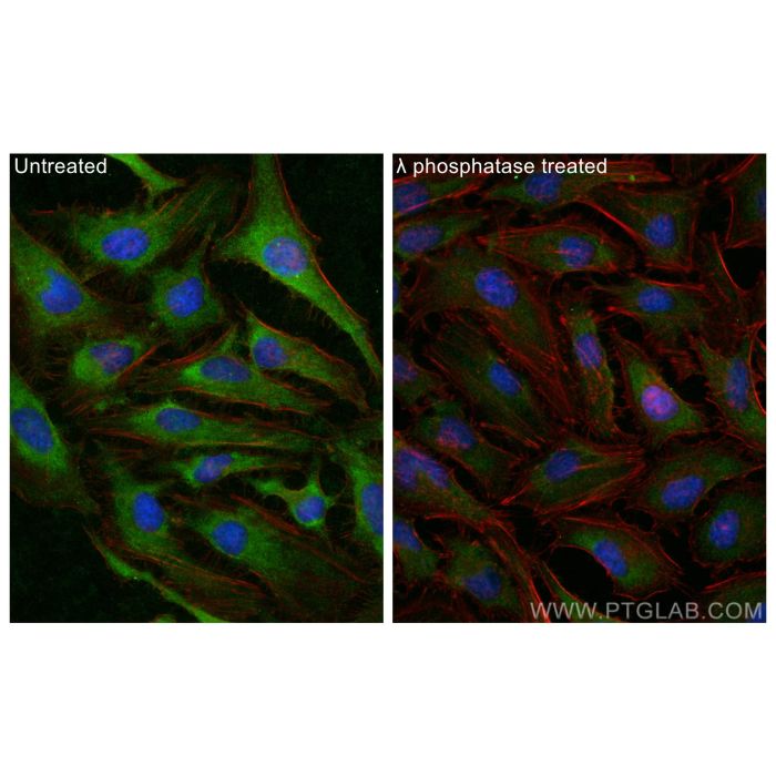
| Tested Applications — Positive IF/ICC detected in | HeLa cells |
| Recommended Dilutions — Western Blot (WB) | WB : 1:2000-1:10000 |
| Recommended Dilutions — Immunofluorescence (IF)/ICC | IF/ICC : 1:200-1:800 |
| Positive WB detected in | HeLa cells |
| Positive IF/ICC detected in | HeLa cells |
| Western Blot (WB) | WB : 1:2000-1:10000 |
| Immunofluorescence (IF)/ICC | IF/ICC : 1:200-1:800 |
| Tested Reactivity | human |
| Cited Reactivity | human, mouse |
| Class | Polyclonal |
| Type | Antibody |
| Immunogen | Peptide Predict reactive species |
| Full Name | myristoylated alanine-rich protein kinase C substrate |
| Calculated Molecular Weight | 32 kDa |
| Observed Molecular Weight | 80 kDa |
| GenBank Accession Number | BC089040 |
| Gene Symbol | MARCKS |
| Gene ID (NCBI) | 4082 |
| RRID | AB_2923590 |
| Conjugate | Unconjugated |
| Purification Method | Antigen affinity purification |
| UNIPROT ID | P29966 |
| Storage Buffer | PBS with 0.02% sodium azide and 50% glycerol, pH 7.3. |
| Storage Conditions | Store at -20°C. Stable for one year after shipment. Aliquoting is unnecessary for -20 o C storage. 20ul sizes contain 0.1% BSA. |
| IF protocol for Phospho-MARCKS (Ser167/170) antibody 29145-1-AP | Download protocol |
| WB protocol for Phospho-MARCKS (Ser167/170) antibody 29145-1-AP | Download protocol |
| mouse,human | WB Life Sci Alliance The nucleocytoplasmic translocation of HINT1 regulates the maturation of cell density. Authors - Xiaofang Zhang View Article |
| mouse | WB Wound Repair Regen Dendritic Cell-Associated MARCKSL1 Regulates Fibroblast Differentiation During Wound Healing. Authors - Kento Takaya View Article |
| Citations | 2 |
| Dilutions | WB : 1:2000-1:10000 IF/ICC : 1:200-1:800 |
| Isotype | IgG |
| Clonality | Polyclonal |
| Clone Number | - |
| Conjugation | Unconjugated |
The Myristoylated Alanine Rich C-Kinase Substrate (MARCKS) is a ubiquitous, highly conserved protein among vertebrates, which is essential for postnatal survival, and has been widely studied for its functions in the brain and nervous system. Being highly expressed in nervous tissue, particularly during early development but persisting in the adult, it plays numerous roles related to brain growth, neuronal migration, neurite outgrowth, neurotransmitter release, and synaptic plasticity. Protein kinase C (PKC) phosphorylates MARCKS, which converts MARCKS from a membrane-bound protein to a cytoplasmic protein. The phosphorylation site of MARCKS protein is called the effector domain (ED). Its structure is highly conserved. It can be combined with cell membrane, PKC, calcium/calmodulin-dependent kineses (CaMK) and F-actin. Studies have shown that increased membrane-bound, non-phosphorylated MARCKS might be conducive to the stabilization of synaptic morphology. Phosphorylated MARCKS protein (P-MARCKS) can regulate the stability of actin network and alter the synaptic structure. (PMID: 30655546, PMID: 30155805) Protocols Product Specific Protocols IF protocol for Phospho-MARCKS (Ser167/170) antibody 29145-1-AP Download protocol WB protocol for Phospho-MARCKS (Ser167/170) antibody 29145-1-AP Download protocol Standard Protocols Click here to view our Standard Protocols
Publications