GRP78/BIP Polyclonal antibody 11587-1-AP proteintech
$149.00
In stock
SKU
11587-1-AP
| Catalog Number | 11587-1-AP |
| Synonyms | BIP, GRP78, HSPA5, 78 kDa glucose-regulated protein, Binding-immunoglobulin protein |
| Host | Rabbit |
| Reactivity | human, mouse, rat and More (7) |
| Reactivity | human, mouse, rat, chicken, goat, sheep, c.elegans, deer, carp, medaka embryos |
| Form | Liquid |
| Formulation | PBS, Azide, Glycerol |
| Applications | WB, IHC, IF/ICC, FC (Intra), IP, CoIP, ELISA |
| Applications | WB, IHC, IF/ICC, FC (Intra), IP, ELISA |
| Host / Isotype | Rabbit / IgG |
| Tested Applications — Positive WB detected in | BxPC-3 cells, Caco-2 cells, HEK-293 cells, HeLa cells, HepG2 cells, mouse brain tissue, mouse liver tissue, rat brain tissue, rat liver tissue, HT-29 cells |
| Tested Applications — Positive IP detected in | MCF-7 cells |
| Tested Applications — Positive IHC detected in | human pancreas cancer tissue, human breast cancer tissue, mouse kidney tissue, rat brain tissue, mouse heart tissue, human colon cancer tissue Note: suggested antigen retrieval with TE buffer pH 9.0; (*) Alternatively, antigen retrieval may be performed with citrate buffer pH 6.0 |
| Tested Applications — Positive IF/ICC detected in | HepG2 cells, Thapsigargin treated HepG2 cells |
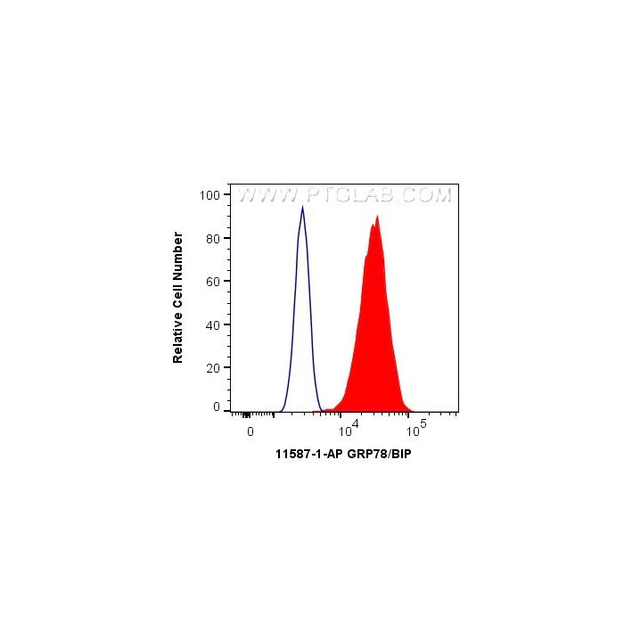
| Tested Applications — Positive FC (Intra) detected in | HeLa cells |
| Recommended Dilutions — Western Blot (WB) | WB : 1:2000-1:12000 |
| Recommended Dilutions — Immunoprecipitation (IP) | IP : 0.5-4.0 ug for 1.0-3.0 mg of total protein lysate |
| Recommended Dilutions — Immunohistochemistry (IHC) | IHC : 1:50-1:500 |
| Recommended Dilutions — Immunofluorescence (IF)/ICC | IF/ICC : 1:50-1:500 |
| Recommended Dilutions — Flow Cytometry (FC) (INTRA) | FC (INTRA) : 0.40 ug per 10^6 cells in a 100 µl suspension |
| Positive WB detected in | BxPC-3 cells, Caco-2 cells, HEK-293 cells, HeLa cells, HepG2 cells, mouse brain tissue, mouse liver tissue, rat brain tissue, rat liver tissue, HT-29 cells |
| Positive IP detected in | MCF-7 cells |
| Positive IHC detected in | human pancreas cancer tissue, human breast cancer tissue, mouse kidney tissue, rat brain tissue, mouse heart tissue, human colon cancer tissue Note: suggested antigen retrieval with TE buffer pH 9.0; (*) Alternatively, antigen retrieval may be performed with citrate buffer pH 6.0 |
| Positive IF/ICC detected in | HepG2 cells, Thapsigargin treated HepG2 cells |
| Positive FC (Intra) detected in | HeLa cells |
| Western Blot (WB) | WB : 1:2000-1:12000 |
| Immunoprecipitation (IP) | IP : 0.5-4.0 ug for 1.0-3.0 mg of total protein lysate |
| Immunohistochemistry (IHC) | IHC : 1:50-1:500 |
| Immunofluorescence (IF)/ICC | IF/ICC : 1:50-1:500 |
| Flow Cytometry (FC) (INTRA) | FC (INTRA) : 0.40 ug per 10^6 cells in a 100 µl suspension |
| Tested Reactivity | human, mouse, rat |
| Cited Reactivity | human, mouse, rat, chicken, goat, sheep, c.elegans, deer, carp, medaka embryos |
| Class | Polyclonal |
| Type | Antibody |
| Immunogen | CatNo: Ag2188 Product name: Recombinant human GRP78,BIP protein Source: e coli. -derived, PGEX-4T Tag: GST Domain: 351-654 aa of BC020235 Sequence: LKKSDIDEIVLVGGSTRIPKIQQLVKEFFNGKEPSRGINPDEAVAYGAAVQAGVLSGDQDTGDLVLLDVCPLTLGIETVGGVMTKLIPRNTVVPTKKSQIFSTASDNQPTVTIKVYEGERPLTKDNHLLGTFDLTGIPPAPRGVPQIEVTFEIDVNGILRVTAEDKGTGNKNKITITNDQNRLTPEEIERMVNDAEKFAEEDKKLKERIDTRNELESYAYSLKNQIGDKEKLGGKLSSEDKETMEKAVEEKIEWLESHQDADIEDFKAKKKELEEIVQPIISKLYGSAGPPPTGEEDTAEKDEL Predict reactive species |
| Full Name | heat shock 70kDa protein 5 (glucose-regulated protein, 78kDa) |
| Calculated Molecular Weight | 78 kDa |
| Observed Molecular Weight | 70-78 kDa |
| GenBank Accession Number | BC020235 |
| Gene Symbol | GRP78/BIP |
| Gene ID (NCBI) | 3309 |
| RRID | AB_2119855 |
| Conjugate | Unconjugated |
| Purification Method | Antigen affinity purification |
| UNIPROT ID | P11021 |
| Storage Buffer | PBS with 0.02% sodium azide and 50% glycerol, pH 7.3. |
| Storage Conditions | Store at -20°C. Stable for one year after shipment. Aliquoting is unnecessary for -20 o C storage. 20ul sizes contain 0.1% BSA. |
| FC protocol for GRP78/BIP antibody 11587-1-AP | Download protocol |
| IF protocol for GRP78/BIP antibody 11587-1-AP | Download protocol |
| IHC protocol for GRP78/BIP antibody 11587-1-AP | Download protocol |
| IP protocol for GRP78/BIP antibody 11587-1-AP | Download protocol |
| WB protocol for GRP78/BIP antibody 11587-1-AP | Download protocol |
| mouse | WB Crit Care Recombinant ACE2 protein protects against acute lung injury induced by SARS-CoV-2 spike RBD protein. Authors - Lingbing Zhang View Article |
| rat,human | WB Acta Pharm Sin B SMYD3-PARP16 axis accelerates unfolded protein response and mediates neointima formation. Authors - Fen Long View Article |
| Hua (Verified Customer) (07-08-2026) | Great antibody for WB. Applications: Western Blot Primary Antibody Dilution: 1:4000 Cell Tissue Type: HepG2, mouse liver and mouse brain. GRP78/BIP Antibody Western Blot validation (1:4000 dilution) in HepG2, mouse liver and mouse brain. (Cat no:11587-1-AP) |
| Melina (Verified Customer) (04-03-2026) | Worked well for western of HEK293T cell lysates Applications: Western Blot Primary Antibody Dilution: 1:1000 Cell Tissue Type: HEK293T cells GRP78/BIP Antibody Western Blot validation (1:1000 dilution) in HEK293T cells (Cat no:11587-1-AP) |
| PK (Verified Customer) (03-22-2022) | Applications: Western Blot Cell Tissue Type: FIBROBLAST GRP78/BIP Antibody Western Blot validation ( dilution) in FIBROBLAST (Cat no:11587-1-AP) |
| Yeawon (Verified Customer) (03-17-2022) | Works very good in mouse kidney tissue. Applications: Western Blot, Immunofluorescence Primary Antibody Dilution: 1:2000 Cell Tissue Type: Mouse kidney tissue |
| Emma (Verified Customer) (03-15-2022) | Works well by WB in LNCaP and CWR22Rv1 cells @ 1:1000. A single band observed. Applications: Western Blot Primary Antibody Dilution: 1:1000 Cell Tissue Type: Prostate |
| P (Verified Customer) (05-27-2021) | Excellent. Applications: Western Blot Primary Antibody Dilution: 1:2000 Cell Tissue Type: H9C2 GRP78/BIP Antibody Western Blot validation (1:2000 dilution) in H9C2 (Cat no:11587-1-AP) |
| Kevin (Verified Customer) (01-24-2019) | Works pretty good in HepG2 cells. One single band was observed. Applications: Western Blot, Primary Antibody Dilution: 1:1000 Cell Tissue Type: HepG2 |
| Sylvain (Verified Customer) (12-03-2018) | Applications: Immunofluorescence, Primary Antibody Dilution: 1:300 Cell Tissue Type: colon cancer cell lines |
| Natalie (Verified Customer) (11-20-2018) | Applications: Western Blot, Primary Antibody Dilution: 1:5000 Cell Tissue Type: Rat Dorsal Root Ganglia GRP78/BIP Antibody Western Blot, validation (1:5000 dilution) in Rat Dorsal Root Ganglia (Cat no:11587-1-AP) |
| Citations | 945 |
| Dilutions | WB : 1:2000-1:12000 IP : 0.5-4.0 ug for IP and 0.5-4.0 ug for 1.0-3.0 mg of total protein lysate for WB IHC : 1:50-1:500 IF/ICC : 1:50-1:500 FC (INTRA) : 0.40 ug per 10^6 cells in a 100 µl suspension |
| Isotype | IgG |
| Clonality | Polyclonal |
| Clone Number | - |
| Conjugation | Unconjugated |
GRP78 (HSPA5), also referred to as 'immunoglobulin heavy chain-binding protein' (BiP), is a member of the heat-shock protein-70 (HSP70) family and is involved in the folding and assembly of proteins in the endoplasmic reticulum (ER). It is a constitutively expressed resident protein of the ER in all eukaryotic cells. Recently it has been reported that GRP78 is associated with apoptosis or inhibition of cancer cell growth. 11587-1-AP antibody recognizes one or two bands with MW 70-78 kDa for the signal peptide of GRP78 protein can be finally cleaved. Protocols Product Specific Protocols FC protocol for GRP78/BIP antibody 11587-1-AP Download protocol IF protocol for GRP78/BIP antibody 11587-1-AP Download protocol IHC protocol for GRP78/BIP antibody 11587-1-AP Download protocol IP protocol for GRP78/BIP antibody 11587-1-AP Download protocol WB protocol for GRP78/BIP antibody 11587-1-AP Download protocol Standard Protocols Click here to view our Standard Protocols
Publications