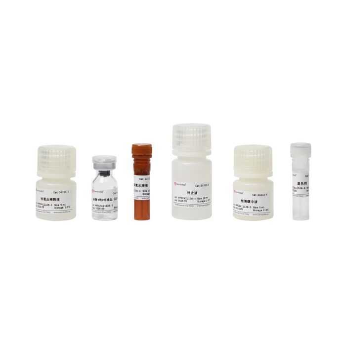
Glutathione Peroxidase (GSH-PX) Assay kit servicebio G4310-48T
$50.00
In stock
SKU
G4310-48T
Product Information
| Product Name | Cat. No. |
| Spec. | Glutathione Peroxidase (GSH-PX) assay kit |
| G4310-48T | 48T |
| G4310-96T | 96T |
Product Description/Introduction
Glutathione peroxidase (GSH-Px, GPX) is an important peroxide-breaking enzyme that exists widely in the body. The active center of GSH-Px is selenocysteine, and its activity can reflect the level of selenium (Se) in the body. GPX can catalyze GSH to GSSG, reduce toxic peroxides to non-toxic hydroxyl compounds, and promote the decomposition of H2O2, so as to protect the structure and function of the cell membrane from interference and damage by oxides.
This product detects glutathione peroxidase containing selenium. The principle is that GPX catalyzes H2O2 oxidation of GSH to produce GSSG, while GSH can react with the chromogenic substrate DTNB to generate GSSG and yellow TNB, then the activity of GPX can be reflected by the decrease of absorbance at 412 nm (GSH consumption). Since these two substrates can also undergo REDOX reactions in the absence of enzymes (called non-enzymatic reactions), the reduction of GSH caused by non-enzymatic reactions must be deducted in the final calculation of the enzyme activity.
2GSH+H2O2+GSH-PX → GSSG+2H2O
2GSH+DTNB → GSSG+2TNB
Storage and Shipping Conditions
Store the kit at 2-8°C for 6 months.
Product Content
| Component Number | Component |
| G4310-50T | G4310-100T |
| G4310-1 | Standard Diluent |
| 5 mL | 10 mL |
| G4310-2 | Glutathione Standard (GSH powder) |
| 1 tube | 2 tube |
| G4310-3 | Hydrogen Peroxide Solution |
| 100 μL | 200 μL |
| G4310-4 | Stop Solution |
| 10 mL | 20 mL |
| G4310-5 | Detection Buffer |
| 5 mL | 10 mL |
| G4310-6 | Developer Solution |
| 30 μL | 60 μL |
| Manual | One copy |
Assay Protocol / Procedures
1. Sample/reagent preparation:
1.1. Dissolved blood, serum (plasma) samples: according to the inhibition rate of the pre-test, select the appropriate dilution times for detection;
1.2. Tissue sample: Homogenate cracking with PBS, where the ratio of tissue to homogenate is 1 to 9 (e.g. 0.1g tissue to 0.9 mL homogenate); After homogenate cracking, centrifuge at 4℃ at 10000 g for 10-15 min, take the supernatant and put it on the ice to be measured. The remaining supernatant was used for protein concentration determination.
1.3. Cell samples: After cell collection, approximately every 1×10^6 cells were homogenized with 300-500 μL PBS; After homogenate cracking, centrifuge at 4℃ at 10000 g for 10-15 min, take the supernatant and put it on the ice to be measured. The remaining supernatant was used for protein concentration determination.
1.4. 1 mM GSH preparation: Centrifuge Glutathione Standard (GSH powder) at low speed to ensure that the powder is at the bottom of the tube; 1 mmol/L reduced glutathione standard stock solution was obtained by using 2 mL Standard Diluent. It is recommended to store at -20℃ after packaging and use up within 1 month.
1.5. Hydrogen peroxide working solution: Hydrogen Peroxide Solution, use sterile ultra-pure water 100 times dilution, ready for use.
1.6. Developer working solution: Developer Solution, use sterile ultra-pure water 100 times dilution, ready for use.
2. GSH-PX test:
2.1. Pre-experiment:
During sample detection, it is necessary to carry out dilution treatment, so that the detection of GSH-Px inhibition rate ranges from 20%-55%, 25%-50% is the best, and the corresponding sample concentration is the best sampling concentration. Therefore, it is necessary to dilute the sample according to the actual situation after the pre-experiment. Maximum sampling concentration search:
Measurement of whole blood: Take 20 μL of hemolytic solution diluted with distilled water at 1:24, 1:49, 1:99, and 1:149, and perform the pre-test according to the formal experimental procedure table;
Serum measurement: Dilute the serum with physiological saline at a ratio of 1:1, 1:4, 1:9, and 1:19, taking 20 μL each, follow the formal experimental procedure table for pre-testing;
Measurement of tissue homogenization: After homogenization, the sample is diluted 0, 2, 5, and 10 times with homogenization solution, and 20 μL of each is taken for pre testing according to the formal experimental operation table;
Calculation of inhibition rate: Inhibition rate=(Non enzyme tube OD value-enzyme tube OD value)/Non enzyme tube OD value ×100%
If the inhibition rate is greater than 60%, the sample concentration needs to be diluted again before testing. If the inhibition rate is less than 20%, the sample concentration needs to be increased or the sample size needs to be increased.
2.2. Formal experiments:
Refer to the table below, add each solution in sequence and mix well. It should be noted that there are significant differences in the activity levels of glutathione peroxidase in samples from different cells or tissues. Before formal testing, 2-3 samples with significant expected differences need to be diluted to different concentrations, and dilution multiples with inhibition rates of 20% -55% (refer to the preliminary experiment) should be selected for formal batch experiments.
2.2.1 Enzymatic reaction:
37℃ water bath, pre heated for 5 minutes
37℃ water bath, accurate reaction for 5 minutes
4000 r/min, centrifuge for 10 minutes. Take 100 μL of supernatant for color development reaction.
| Enzyme tube | Non enzymatic tube |
| Sample | 20 μL |
| 1 mM GSH | 20 μL |
| 20 μL | Hydrogen peroxide working solution |
| 10 μL | 10 μL |
| Stop Solution | 200 μL |
| 200 μL | Sample |
| 20 μL |
2.2.2 Color development reaction:
React at 37 ℃ for 5 minutes and read the absorbance value A at a wavelength of 412 nm.
| Enzyme tube | Non enzymatic tube |
| Blank tubec | Standard tubec |
| Enzymatic reaction supernatant | 100 μL |
| 100 μL | Blank Reagenta |
| 100 μL | 20 μM GSH Standard Solutionb |
| 100 μL | Detection Buffer |
| 100 μL | 100 μL |
| 100 μL | 100 μL |
| Developer working solution | 50 μL |
| 50 μL | 50 μL |
| 50 μL |
Note a: Blank Reagent 100 μL: 20 μL standard dilution solution+80 μL Stop Solution.
Note b: 20 μM GSH standard solution: 1 mmol/L GSH standard storage solution, diluted 10 times with standard diluent to obtain 100 μmol/L standard solution. Take 20 μL of 100 μmol/L standard solution and 80 μL of Stop Solution to obtain 100 μL of 20 μ M GSH standard solution.
Note c: Blank tubes and Standard tubes generally only need to be made in 1-2 tubes. The sample needs to be tested as soon as possible after fragmentation, as long-term storage may lead to decreased vitality.
3. Enzyme activity calculation:
3.1. Organizational and cellular samples:
Definition: Deducting the effect of non enzymatic reactions, the catalytic activity of 1 nmol GSH oxidation per minute per milligram of protein in the reaction system is defined as one enzyme activity unit.
Calculation formula:
GPX vitality (U/mg prot)=(non enzyme tube OD value-enzyme tube OD value)/(standard tube OD value-blank tube OD value)×Cstandard×V1/(V2×Cpr)/T×f
Note: Cstandard: the standard tube concentration is 20 μmol/L; V1: enzymatic reaction volume 0.25 mL; V2: sample volume added in the reaction, 0.02 mL; Cpr: sample protein concentration, in mg/mL; T: reaction time, 5min; f: dilution ratio of the sample before sample addition
3.2. Calculation of enzyme activity in whole blood and serum (plasma) samples:
Definition: Deducting non enzymatic reactions, catalytic oxidation of 1 nmol GSH per minute per mL of blood sample in the reaction system is defined as one enzyme activity unit.
Calculation formula:
GPX vitality (U/mL)=(Non enzyme tube OD value-enzyme tube OD value)/(standard tube OD value - blank tube OD value)×Cstandard×V1/V2/T×f
Note: Cstandard: the standard tube concentration is 20 μmol/L; V1: enzymatic reaction volume 0.25 mL; V2: sample volume added in the reaction, 0.02 mL; T: reaction time, 5min; f: dilution ratio of the sample before sample addition.
4. Preparation of GSH standard curve
4.1. Standard Dilution: Dilute 1 mmol/L GSH standard storage solution with standard dilution to 400, 200, 100, 50, 25, 12.5, 6.25, and 0 μmol/L. Take 20 μL/tube of each dilution solution, add 80 μL of Stop Solution for dilution, and obtain standard concentration of 0, 1.25, 2.5, 5, 10, 20, 40, and 80 μmol/L.
4.2. Color development: Add 100 μL of detection buffer and 50 μL of color development agent to the above-mentioned tubes, react at 37 ℃ for 1 minute, and read the absorbance value A at a wavelength of 412 nm.
4.3. Drawing: Draw the standard curve with GSH standard concentration as the x-axis and absolute value OD (minus blank) as the y-axis.
Note
1. The detection of this reagent kit involves redox reactions, so all oxidants or reducing agents will interfere with the determination of this reagent kit. If the reducing agents in the sample cannot be avoided, such as DTT, mercaptoethanol, etc., the total concentration of these reducing agents is at least less than 0.1 mM. 0.15 mM DTT can inhibit 40% of enzyme activity.
2. Commonly used descaling agents such as Triton X-100 and Tween-20 contain high levels of peroxides, which can affect the determination of this reagent kit. If it is necessary to use these descaling agents, it is best to use descaling agents with higher purity and indicating lower peroxide content.
3. The BCA protein quantitative detection kit (G2026) can be used for sample protein concentration determination.
4. For your health and safety, please wear laboratory clothes and gloves when operating.
Appendix: Technical parameters
| Detection index | Glutathione Peroxidase (GSH-PX) |
| Sample type | blood, tissue, cell |
| Detection Sensitivity and Detection Range | 1.25 μmol/L;1.25-80 μmol/L |
| Recovery Rate | 100±5% |
| Intra-batch CV | <3% |
| Inter-batch CV | <5% |
For Research Use Only!