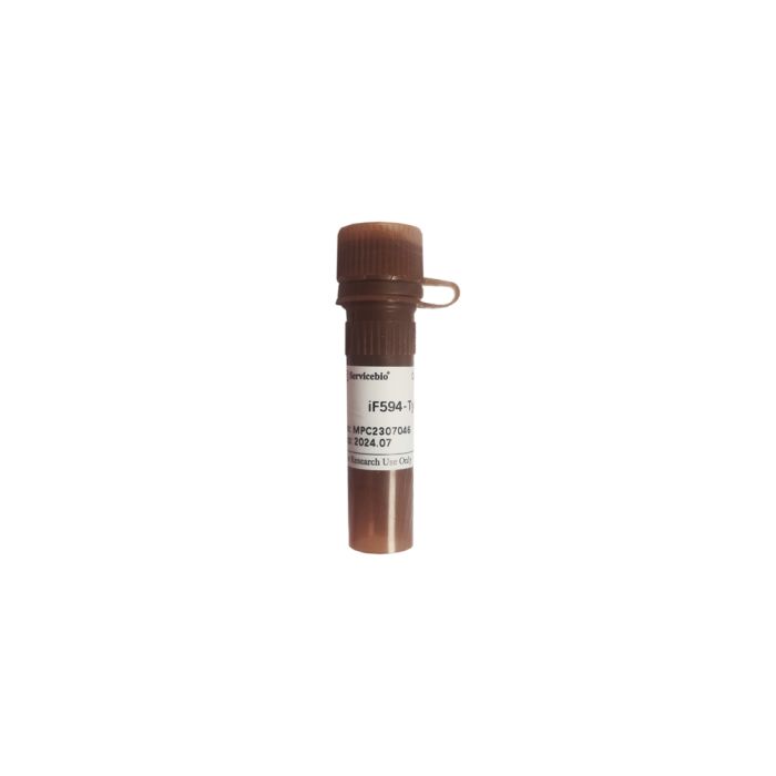
iF594-Tyramide servicebio G1242-25UL
$50.00
In stock
SKU
G1242-25UL
Product Information
| Product Name: iF 594 -Tyramide | Cat.No.: G12 42 -50UL |
| Spec.: 50 μL |
Description
This product, iF594-Tyramide, is a tyramine labeled with fluorescent dye IF594. Tyramide signal amplification (TSA) is a type of enzymatic detection method that uses HRP to perform high-density in-situ labeling of target antigens and can be applied to signal amplification of IF/IHC. Main principles of TSA technology: In hydrogen peroxide H2O2, horseradish peroxidase (HRP) can catalytic tyramine for a very short life of active intermediates, in a short period of time, intermediates can be covalent bonding to the surrounding protein rich electronic surface tyramine chemical compound, a large number of rich integration with the center of the enzyme poly group, such as the "island", by using the fluorescent dye iF594 tyramine, the antigen-antibody binding site to form a large amount of fluorescein deposition, to achieve signal amplification. In addition, multiple fluorescent staining can be achieved by repeated immunolabeling several times using different fluorescent tyramines.
The fluorescence spectrum data of the series products are as follows:
| Product Code: G1222-50UL | Product Name: FITC-Tyramide |
| Spec.: 50 μ L | Ex/Em: 492/518 |
| Product Code: G1223-50UL | Product Name: CY3-Tyramide |
| Spec.: 50 μ L | Ex/Em: 555/569 |
| Product Code: G1231-50UL | Product Name: iF488-Tyramide |
| Spec.: 50 μ L | Ex/Em: 491/516 |
| Product Code: G1233-50UL | Product Name: iF555-Tyramide |
| Spec.: 50 μ L | Ex/Em: 557/570 |
| Product Code: G12 42 -50UL | Product Name: iF 594 -Tyramide |
| Spec.: 50 μ L | Ex/Em: 588/604 |
| Product Code: G1232-50UL | Product Name: iF647-Tyramide |
| Spec.: 50 μ L | Ex/Em: 656/670 |
Storage and Handling Conditions
Wet ice packs for transportation, store at -20℃ for 12 months.
Component
Product Manual
| Component: iF 594 -Tyramide | G12 42 -50UL: 50 μ L |
Assay Protocol
Take tissue sections as an example. The water used in the following experimental steps is pure water, and the reagents involved should be prepared by users themselves, or refer to Servicebio related products:
1. Dewaxed tissue sections to water;
2. Antigen repair: According to the primary antibody used and the type of sample, appropriate methods were used to repair the antigen of tissue sections. Wash with 1×PBS (G4202 or G0002 is recommended) three times, 5 min each time;
3. Use immunohistochemical pen to mark tissues in circles;
4. (Optional) Add tissue autofluorescence quencher (G1221A solution is recommended) to the tissue, incubate at room temperature for 30 min, and wash with water for 5 min;
5. Inactivation of endogenous peroxidase: 3% H2O2 was added to the section to cover the tissue and incubated at room temperature in the dark for 25 min to block endogenous peroxidase and reduce non-specific background staining. Wash with 1×PBS 3 times, 5 min each time;
6. Block: After the sections are slightly dried with water, block them with 3%BSA or serum for 30 min. If the primary antibody was of goat origin, it was blocked with 10% donkey serum and other sources were blocked with 3%BSA.
7. Incubation of primary antibody: The primary antibody was diluted to the appropriate concentration with 1×PBS or blocking solution (G2010 recommended). The blocking solution on the section was gently removed, and the diluted primary antibody was added to cover the tissue. The section was placed flat in a wet box with water and incubated overnight at 4℃. Wash with 1×PBS 3 times, 5 min each time;
8. Incubation of HRP-labeled secondary antibodies: The secondary antibodies were diluted to an appropriate concentration with 1×PBS or blocking solution (G2010 recommended), and the secondary antibodies were added to the tissues by drop, and incubated for 50 min at room temperature. Wash with 1×PBS 3 times, 5 min each time;
9. Prepare Tyramide diluent containing 0.03‰H2O2: 100 mL 1×TBST (recommended G0004), add 100 μL 3% H2O2, and mix well. Because H2O2 is easy to decompose, this solution needs to be prepared for use.
10. Preparation of TSA staining working solution: The Tyramide dilution obtained in the previous step was diluted 500 times with iF594-Tyramide. To obtain TSA staining working solution, every 1 mL Tyramide dilution was mixed with 2 μL iF594-Tyramide. Add 100 μL staining solution to each sample, and incubate at room temperature for 10 min under light. Wash with 1×PBS 3 times, 5 min each time;
(Note: TSA staining solution is recommended to be prepared and used now. It should be stored at 4℃ and protected from light for 24 hours.)
11. Microwave treatment: Tissue sections were placed in a repair box filled with antigen repair solution (appropriate antigen repair solution selected according to the tissue type and antibody) for microwave heating treatment to remove the bound primary and secondary antibodies. During this step, it is necessary to prevent excessive evaporation of liquid resulting in dry flakes;
(Note: If double or multiple fluorescent staining is required, proceed as follows. If double or multiple fluorescent staining is not required, skip this step and proceed to Step 12)
12. Repeat steps 7-11 to incubate the second primary antibody, secondary antibody, and TSA labeling, and remove the primary antibody and secondary antibody by microwave treatment;
13. Nuclear counterstaining DAPI: After the sections were slightly dried, DAPI staining solution (G1012 is recommended) was added to the tissues, and the cells were incubated for 10 min at room temperature in the dark. Wash with 1×PBS for 3 times, 5 min each time.
14. (Optional) Tissue autofluorescence quenching: Add autofluorescence quenching agent (liquid G1221B recommended) to the tissue, incubate at room temperature for 5 min, and rinse with running water for 3 min.
15. Sealing and microscopic examination: After the sections are slightly dried, drop the anti-fluorescence quenched tablet to seal the slices (G1401 is recommended). Select appropriate fluorescence channel to observe and capture images. The slices can be stored at 4℃ for 15 days in a light protected box.
Note:
1. After the product is melted at room temperature, centrifuge it briefly, blow and mix it with a clean suction head, then take it out.
2. Compared with fluorescent secondary antibody, TSA kit has higher sensitivity and stronger signal. Therefore, the concentration of primary antibody should be reduced, and the dilution recommended by the antibody manual should be increased 5-10 times to reduce the background fluorescence caused by non-specific binding. It is recommended to set the gradient concentration of primary antibody to obtain the best effect.
3. If the background fluorescence is strong, add tissue autofluorescence quenching step.
4. The recommended dilution ratio of fluorescent Tyramide is 1:500, and the dilution ratio can be adjusted according to the experimental results (the recommended dilution ratio ranges from 1:200 to 1:1000).
5. If multiple fluorescent labeling is performed, it is recommended to incubate the polyclonal antibody first and then the monoclonal antibody; the antibody corresponding to the high abundance target protein was incubated first, and then the antibody corresponding to the low abundance target protein was incubated.
6. Wear a lab coat and disposable gloves during operation.
For Research Use Only!