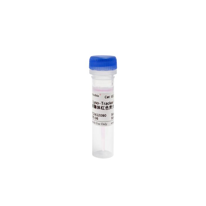
Lyso-Tracker Red servicebio G1741-50UL
$50.00
In stock
SKU
G1741-50UL
Product Information
| Product Name | Cat. No. |
| Spec. | Lyso-Tracker Red |
| G1741-50UL | 50 μL |
Product Description/Introduction
Lyso-Tracker Red DND-99 is a fluorescently labeled red fluorescent probe with a weak base, in which only the weak base partially provides protons to maintain pH neutrality and selectively retains them in acidic lysosomes for specific fluorescent labeling of lysosomes. The maximum excitation and emission wavelengths of Lyso-Tracker Red DND-99 are 577 nm/590 nm, which can be effectively excited using TRITC filter sets. Although the Lyso-Tracker probe does not cross-link to cells, Lyso-Tracker Red DND-99 can be retained transiently after a short period of time after glutaraldehyde fixation.
Storage and Shipping Conditions
Ship with dry ice; Store at -20℃ away from light for 6months.
Product Content
| Component | G1741-50UL |
| Lyso-Tracker Red | 50 μL |
| Manual | 1 pc |
Assay Protocol / Procedures
1. Assay working solution preparation:
1.1. This product is a 1 mM storage solution. Thaw to room temperature before use, perform a brief low-speed centrifugation to ensure that the reagent settles at the bottom of the tube;
1.2. It is recommended to use serum-free medium (Hanks, PBS, etc. can also be used) to dilute the probe into a lysosomal assay working solution of 50-75 nM (50 nM has been tested to be sufficient to stain cells;
2. Cell staining (the following protocol is for adherent cells; centrifugation is required for suspension cell exchange)
2.1. Normal or treated cells should be washed 2-3 times with serum-free medium;
2.2. Add pre-warmed lysosomal assay working solution and incubate at 37℃ away from light for 30 min (the concentration of assay solution and incubation time can be adjusted for different cells);
2.3. Remove the probe and wash 2-3 times with serum-free medium;
3. Microscopy: Observe under a fluorescence microscope; excitation can be achieved using the DsRed channel.
Note
1. Kinetic studies of the endocytosis of Lyso-Tracker Red DND-99 probe show that the dye can be taken up by live cells in just a few seconds. However, these lysosomal probes can lead to the alkalinization of lysosomes, and prolonged incubation can cause an increase in lysosomal pH. Therefore, it is recommended that the incubation time with the probe before imaging should not be too long.
2. This dye is suitable for living cells and cannot be stained after fixation. After staining, it can be fixed with glutaraldehyde, but it will slightly reduce the fluorescence intensity.
3. Fluorescent dyes have quenching problems, please try to avoid light to slow down the quenching of fluorescence.
4. It is recommended to use the probe for the first time after dispensing to avoid repeated freezing and thawing.
5. For your health and safety, please wear lab coat and gloves during operation.
For Research Use Only!