
Anti-CD4 Antibody, Rabbit Polyclonal sinobiological 10400-T24
$243.00
In stock
SKU
10400-T24
| Catalog No. | Size | Price (USD) |
| 10400-T24-100 | 100 µL | $251.64 |
| Catalog No. | Size | Price (USD) |
| 10400-T24-100 | 100 µL | $251.64 |
General Information
| Product name | Anti-CD4 Antibody, Rabbit Polyclonal |
| Validated applications | ELISA (FAQ Protocol), IHC-P (FAQ Protocol) |
| Species reactivity | Reacts with: Human |
| Specificity | Human CD4 |
| Immunogen | Recombinant Human CD4 Protein (Catalog#10400-H08H) |
| Preparation | Produced in rabbits immunized with purified, recombinant Human CD4 (rh CD4; Catalog#10400-H08H; NP_000607.1; Met1-Trp390). CD4 specific IgG was purified by Human CD4 affinity chromatography. |
| Source | Polyclonal Rabbit IgG |
| Purification | Protein A & Antigen Affinity |
| Formulation | 0.2 μm filtered solution in PBS |
| Conjugate | Unconjugated |
| Form | Liquid |
| Shipping | This antibody is shipped as liquid solution at ambient temperature. Upon receipt, store it immediately at the temperature recommended below. |
| Storage | This antibody can be stored at 2℃-8℃ for one month without detectable loss of activity. Antibody products are stable for twelve months from date of receipt when stored at -20℃ to -80℃. Preservative-Free. Avoid repeated freeze-thaw cycles. |
Synonyms: Anti-CD4mut Antibody; Anti-IMD79 Antibody; Anti-Leu-3 Antibody; Anti-OKT4D Antibody; Anti-T4 Antibody
Image / Experimental Information
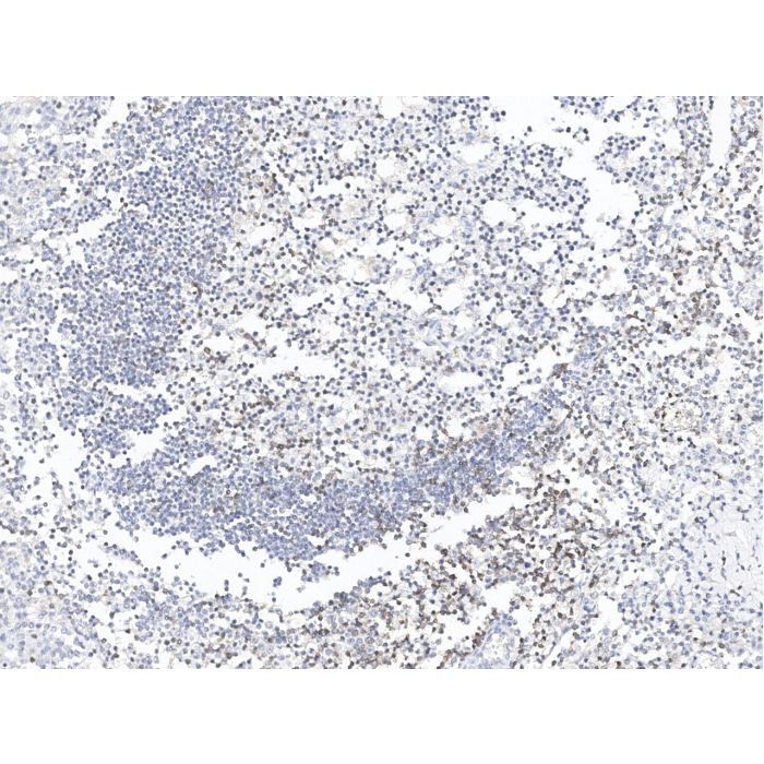
| Image 1 Description | Immunochemical staining of human CD4 in human tonsil with rabbit polyclonal antibody at 1:1000 dilution, formalin-fixed paraffin embedded sections. |
| Image 1 Alt Text | Human CD4 Immunohistochemistry(IHC) 38835 |
| Image 1 URL | Source image |
| Image 2 Description | Immunochemical staining of human CD4 in human tonsil with rabbit polyclonal antibody at 1:1000 dilution, formalin-fixed paraffin embedded sections. |
| Image 2 Alt Text | Human CD4 Immunohistochemistry(IHC) 38836 |
| Image 2 URL | Source image |
Background Information
| Full Name | CD4 molecule |
| Description | T-cell surface glycoprotein CD4, is a single-pass type I membrane protein. CD4 contains three Ig-like C2-type (immunoglobulin-like) domains and one Ig-like V-type (immunoglobulin-like) domain. CD4 is a glycoprotein expressed on the surface of T helper cells, regulatory T cells, monocytes, macrophages, and dendritic cells. The CD4 surface determinant, previously associated as a phenotypic marker for helper/inducer subsets of T lymphocytes, has now been critically identified as the binding/entry protein for human immunodeficiency viruses (HIV). The human CD4 molecule is readily detectable on monocytes, T lymphocytes, and brain tissues. All human tissue sources of CD4 bind radiolabeled gp120 to the same relative degree; however, the murine homologous protein, L3T4, does not bind the HIV envelope protein. CD4 is a co-receptor that assists the T cell receptor (TCR) to activate its T cell following an interaction with an antigen-presenting cell. Using its portion that resides inside the T cell, CD4 amplifies the signal generated by the TCR. CD4 interacts directly with MHC class II molecules on the surface of the antigen-presenting cell via its extracellular domain. The CD4 molecule is currently the object of intense interest and investigation both because of its role in normal T-cell function, and because of its role in HIV infection. CD4 is a primary receptor used by HIV-1 to gain entry into host T cells. HIV infection leads to a progressive reduction of the number of T cells possessing CD4 receptors. Viral protein U (VpU) of HIV-1 plays an important role in downregulation of the main HIV-1 receptor CD4 from the surface of infected cells. Physical binding of VpU to newly synthesized CD4 in the endoplasmic reticulum is an early step in a pathway leading to proteasomal degradation of CD4. Amino acids in both helices found in the cytoplasmic region of VpU in membrane-mimicking detergent micelles experience chemical shift perturbations upon binding to CD4, whereas amino acids between the two helices and at the C-terminus of VpU show no or only small changes, respectively. Paramagnetic spin labels were attached at three sequence positions of a CD4 peptide comprising the transmembrane and cytosolic domains of the receptor. VpU binds to a membrane-proximal region in the cytoplasmic domain of CD4. |
| Related Pathways |
|
| References |
|
title: Gasdermin C promotes Stemness and Immune Evasion in Pancreatic Cancer via Pyroptosis‐Independent Mechanism
authors: Renfei Wu; Jingwei Li; ...; Christopher Heeschen
journal: Advanced Science
date: 2024-09-19
pmid: 39297408
doi: 10.1002/advs.202308990
pmcid: 11558074
cited_by_count: 9
techniques: Cell Fractionation; Control; Derivative Assay; Expressing; Flow Cytometry; Immunofluorescence; Immunohistochemistry; Immunoprecipitation; In Vivo; Inhibition; Injection; Knockdown; MANN-WHITNEY; Marker; Negative Control; Staining; Two Tailed Test; Western Blot
bioz_score: 92
title: T-cell immunoglobulin and mucin 1 (TIM-1) mediates infection of Hantaan virus in Jurkat T cells
authors: Ruixue Ma; Xuyang Zheng; ...; Xingan Wu
journal: Virus research
date: 2024-05-24
doi: 10.1016/j.virusres.2024.199394
pmcid: 11152704
cited_by_count: 2
techniques: Binding Assay; CCK-8 Assay; Expressing; Flow Cytometry; Incubation; Infection; Isolation; Negative Control; Real-time Polymerase Chain Reaction; Staining; Two Tailed Test; Western Blot
bioz_score: 92
-HD17MY2927-1000-human-tonsil-IHC-P.png)
-HD17MY2927-1000-human-spleen-IHC-P.png)