Recombinant Rhesus CD4 Protein (Catalog#90274-C08H)
Preparation
This antibody was produced from a hybridoma resulting from the fusion of a mouse myeloma with B cells obtained from a mouse immunized with purified, recombinant Rhesus CD4 (Catalog#90274-C08H; EHH20435.1; Met1-Trp390) and conjugated with FITC under optimum conditions, the unreacted FITC was removed.
Source
Monoclonal Mouse IgG1 Clone #53
Purification
Protein A
Formulation
PBS solution containing 0.5% BSA and 0.03%ProClin300
Concentration
10 μl/Test, 0.1 mg/ml
Conjugate
FITC
Form
Liquid
Shipping
This antibody is shipped as liquid solution at ambient temperature. Upon receipt, store it immediately at the temperature recommended below.
Storage
This antibody can be stored at 2℃-8℃ for twelve months without detectable loss of activity. Protected from prolonged exposure to light. Do not freeze !
Image / Experimental Information
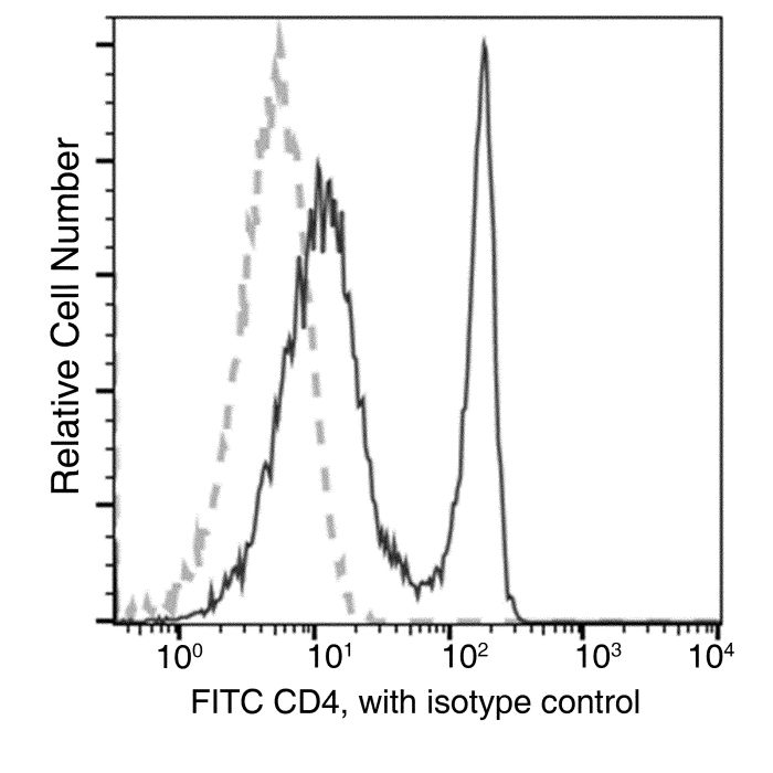
Image 1 Description
Flow cytometric analysis of Cynomolgus CD4 expression on Cynomolgus lymphocytes. Cells were stained with FITC-conjugated anti-CD4. The fluorescence histograms were derived from gated events with the forward and side light-scatter characteristics of viable lymphocytes.
T-cell surface glycoprotein CD4, is a single-pass type I membrane protein. CD4 contains three Ig-like C2-type (immunoglobulin-like) domains and one Ig-like V-type (immunoglobulin-like) domain. CD4 is a glycoprotein expressed on the surface of T helper cells, regulatory T cells, monocytes, macrophages, and dendritic cells. The CD4 surface determinant, previously associated as a phenotypic marker for helper/inducer subsets of T lymphocytes, has now been critically identified as the binding/entry protein for human immunodeficiency viruses (HIV). The human CD4 molecule is readily detectable on monocytes, T lymphocytes, and brain tissues. All human tissue sources of CD4 bind radiolabeled gp120 to the same relative degree; however, the murine homologous protein, L3T4, does not bind the HIV envelope protein. CD4 is a co-receptor that assists the T cell receptor (TCR) to activate its T cell following an interaction with an antigen-presenting cell. Using its portion that resides inside the T cell, CD4 amplifies the signal generated by the TCR. CD4 interacts directly with MHC class II molecules on the surface of the antigen-presenting cell via its extracellular domain. The CD4 molecule is currently the object of intense interest and investigation both because of its role in normal T-cell function, and because of its role in HIV infection. CD4 is a primary receptor used by HIV-1 to gain entry into host T cells. HIV infection leads to a progressive reduction of the number of T cells possessing CD4 receptors. Viral protein U (VpU) of HIV-1 plays an important role in downregulation of the main HIV-1 receptor CD4 from the surface of infected cells. Physical binding of VpU to newly synthesized CD4 in the endoplasmic reticulum is an early step in a pathway leading to proteasomal degradation of CD4. Amino acids in both helices found in the cytoplasmic region of VpU in membrane-mimicking detergent micelles experience chemical shift perturbations upon binding to CD4, whereas amino acids between the two helices and at the C-terminus of VpU show no or only small changes, respectively. Paramagnetic spin labels were attached at three sequence positions of a CD4 peptide comprising the transmembrane and cytosolic domains of the receptor. VpU binds to a membrane-proximal region in the cytoplasmic domain of CD4.
Related Pathways
NF-kB (NFkB) Pathway
T Cell Receptor Signaling Pathway
References
Farrar WL, et al. (1988) Characterization of CD4 glycoprotein determinant-HIV envelope protein interactions: perspectives for analog and vaccine development. Crit Rev Immunol. 8(4): 315-39.
Biddison WE, et al. (1989) CD4 expression and function in HLA class II-specific T cells. Immunol Rev. 109: 5-15.
Singh SK, et al. (2012) Mapping the interaction between the cytoplasmic domains of HIV-1 viral protein U and human CD4 with NMR spectroscopy. FEBS J. 279(19):3705-14.