
Anti-Desmin Antibody, Rabbit Polyclonal sinobiological 106974-T42
$243.00
In stock
SKU
106974-T42
| Catalog No. | Size | Price (USD) |
| 106974-T42-100 | 100 µL | $251.64 |
| Catalog No. | Size | Price (USD) |
| 106974-T42-100 | 100 µL | $251.64 |
General Information
| Product name | Anti-Desmin Antibody, Rabbit Polyclonal |
| Validated applications | WB (FAQ Protocol), IHC-P (FAQ Protocol), ICC/IF (FAQ Protocol) |
| Species reactivity | Reacts with: Human |
| Specificity | Human Mouse, Rat, Bovin, Pig (Species predicted to react based on 100% sequence homology) |
| Immunogen | A synthetic peptide corresponding to the C-terminus of the Human Desmin |
| Preparation | Produced in rabbits immunized with a synthetic peptide corresponding to the C-terminus of the Human Desmin, and purified by antigen affinity chromatography. |
| Source | Polyclonal Rabbit IgG |
| Purification | Protein A & Antigen Affinity |
| Formulation | 0.2 μm filtered solution in PBS |
| Conjugate | Unconjugated |
| Form | Liquid |
| Shipping | This antibody is shipped as liquid solution at ambient temperature. Upon receipt, store it immediately at the temperature recommended below. |
| Storage | This antibody can be stored at 2℃-8℃ for one month without detectable loss of activity. Antibody products are stable for twelve months from date of receipt when stored at -20℃ to -80℃. Preservative-Free. Avoid repeated freeze-thaw cycles. |
Synonyms: Anti-CDCD3 Antibody; Anti-CMD1I Antibody; Anti-CSM1 Antibody; Anti-CSM2 Antibody; Anti-LGMD1D Antibody; Anti-LGMD1E Antibody; Anti-LGMD2R Antibody
Image / Experimental Information
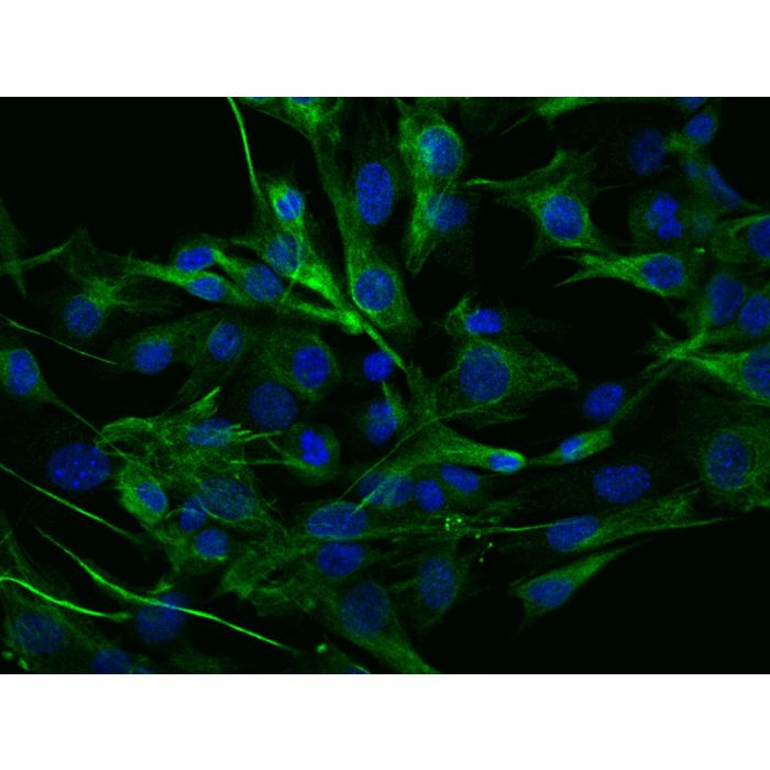
| Image 1 Description | Immunofluorescence staining of Desmin in C2C12 cells. Cells were fixed with 4% PFA, permeabilzed with 0.3% Triton X-100 in PBS,blocked with 10% serum, and incubated with rabbit anti-mouse Desmin polyclonal antibody (dilution ratio 1:100) at 4℃ overnight. Then cells were stained with the Alexa Fluor®488-conjugated Goat Anti-rabbit IgG secondary antibody (green) and counterstained with DAPI (blue).Positive staining was localized to Cytoplasm. |
| Image 1 Alt Text | Human Desmin Immunofluorescence(IF) 36927 |
| Image 1 URL | Source image |
| Image 2 Description | Immunofluorescence staining of Desmin in C2C12 cells. Cells were fixed with 4% PFA, permeabilzed with 0.3% Triton X-100 in PBS,blocked with 10% serum, and incubated with rabbit anti-mouse Desmin polyclonal antibody (dilution ratio 1:100) at 4℃ overnight. Then cells were stained with the Alexa Fluor®488-conjugated Goat Anti-rabbit IgG secondary antibody (green) and counterstained with DAPI (blue).Positive staining was localized to Cytoplasm. |
| Image 2 Alt Text | Human Desmin Immunohistochemistry(IHC) 36926 |
| Image 2 URL | Source image |
| Image 3 Description | Immunofluorescence staining of Desmin in C2C12 cells. Cells were fixed with 4% PFA, permeabilzed with 0.3% Triton X-100 in PBS,blocked with 10% serum, and incubated with rabbit anti-mouse Desmin polyclonal antibody (dilution ratio 1:100) at 4℃ overnight. Then cells were stained with the Alexa Fluor®488-conjugated Goat Anti-rabbit IgG secondary antibody (green) and counterstained with DAPI (blue).Positive staining was localized to Cytoplasm. |
| Image 3 Alt Text | Human Desmin Immunohistochemistry(IHC) 36925 |
| Image 3 URL | Source image |
| Image 4 Description | Immunofluorescence staining of Desmin in C2C12 cells. Cells were fixed with 4% PFA, permeabilzed with 0.3% Triton X-100 in PBS,blocked with 10% serum, and incubated with rabbit anti-mouse Desmin polyclonal antibody (dilution ratio 1:100) at 4℃ overnight. Then cells were stained with the Alexa Fluor®488-conjugated Goat Anti-rabbit IgG secondary antibody (green) and counterstained with DAPI (blue).Positive staining was localized to Cytoplasm. |
| Image 4 Alt Text | Human Desmin Immunohistochemistry(IHC) 36924 |
| Image 4 URL | Source image |
| Image 5 Description | Immunofluorescence staining of Desmin in C2C12 cells. Cells were fixed with 4% PFA, permeabilzed with 0.3% Triton X-100 in PBS,blocked with 10% serum, and incubated with rabbit anti-mouse Desmin polyclonal antibody (dilution ratio 1:100) at 4℃ overnight. Then cells were stained with the Alexa Fluor®488-conjugated Goat Anti-rabbit IgG secondary antibody (green) and counterstained with DAPI (blue).Positive staining was localized to Cytoplasm. |
| Image 5 Alt Text | Human Desmin Immunohistochemistry(IHC) 36923 |
| Image 5 URL | Source image |
Background Information
| Full Name | desmin |
| Description | The cohesin loading factor, nipped-B-like protein (NIPBL), is also known as the sister chromatid cohesion 2 (SCC2) human homolog, is a cohesin loading factor which is essential for deposition of cohesin onto the sister chromatid. Recent studies have shown that NIPBL contributes to sister chromatid cohesion and plays a critical role in development, DNA repair, and gene regulation. Downregulation of NIPBL arrested cells in the G0/G1 phase of the cell cycle and induced apoptosis and autophagy of breast cancer cells through the caspase3 and mammalian target of rapamycin (mTOR) signaling pathways. NIPBL, the sister chromatid cohesion 2 (SCC2) human homolog, is a cohesin loading factor which is essential for deposition of cohesin onto the sister chromatid. Recent studies have shown that NIPBL contributes to sister chromatid cohesion and plays a critical role in development, DNA repair, and gene regulation. |
| Research Areas |
|
-HD16NO1831-100-C2C12-IF.png)
-HD16NO1831-1000-rat-stomach-IHC-P.png)
-HD16NO1831-100-Mouse-stomach-IHC-P.png)
-HD16NO1831-100-rat-heart-IHC-P.png)
-HD16NO1831-100-Mouse-heart-IHC-P.png)