
Anti-H2AFM/HIST1H2AB Antibody, Rabbit Polyclonal sinobiological 101245-T42
$243.00
In stock
SKU
101245-T42
| Catalog No. | Size | Price (USD) |
| 101245-T42-100 | 100 µL | $251.64 |
| Catalog No. | Size | Price (USD) |
| 101245-T42-100 | 100 µL | $251.64 |
General Information
| Product name | Anti-H2AFM/HIST1H2AB Antibody, Rabbit Polyclonal |
| Validated applications | WB (FAQ Protocol), IHC-P (FAQ Protocol), ICC/IF (FAQ Protocol) |
| Species reactivity | Reacts with: Human |
| Specificity | Human Mouse (Species predicted to react based on 100% sequence homology) |
| Immunogen | A synthetic peptide corresponding to the N-terminus of the Human H2AFM/HIST1H2AB |
| Preparation | Produced in rabbits immunized with a synthetic peptide corresponding to the N-terminus of the Human H2AFM/HIST1H2AB, and purified by antigen affinity chromatography. |
| Source | Polyclonal Rabbit IgG |
| Purification | Protein A & Antigen Affinity |
| Formulation | 0.2 μm filtered solution in PBS |
| Conjugate | Unconjugated |
| Form | Liquid |
| Shipping | This antibody is shipped as liquid solution at ambient temperature. Upon receipt, store it immediately at the temperature recommended below. |
| Storage | This antibody can be stored at 2℃-8℃ for one month without detectable loss of activity. Antibody products are stable for twelve months from date of receipt when stored at -20℃ to -80℃. Preservative-Free. Avoid repeated freeze-thaw cycles. |
Image / Experimental Information
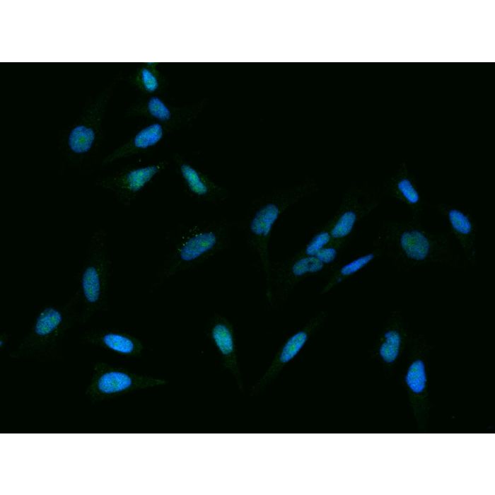
| Image 1 Description | Immunofluorescence staining of H2AFM/HIST1H2AB in Hela cells. Cells were fixed with 4% PFA, permeabilzed with 0.3% Triton X-100 in PBS,blocked with 10% serum, and incubated with rabbit anti-Human H2AFM/HIST1H2AB polyclonal antibody (dilution ratio 1:100) at 4℃ overnight. Then cells were stained with the Alexa Fluor®488-conjugated Goat Anti-rabbit IgG secondary antibody (green) and counterstained with DAPI (blue).Positive staining was localized to Nucleus. |
| Image 1 Alt Text | Human H2AFM/HIST1H2AB Immunofluorescence(IF) 36904 |
| Image 1 URL | Source image |
| Image 2 Description | Immunofluorescence staining of H2AFM/HIST1H2AB in Hela cells. Cells were fixed with 4% PFA, permeabilzed with 0.3% Triton X-100 in PBS,blocked with 10% serum, and incubated with rabbit anti-Human H2AFM/HIST1H2AB polyclonal antibody (dilution ratio 1:100) at 4℃ overnight. Then cells were stained with the Alexa Fluor®488-conjugated Goat Anti-rabbit IgG secondary antibody (green) and counterstained with DAPI (blue).Positive staining was localized to Nucleus. |
| Image 2 Alt Text | Human H2AFM/HIST1H2AB Immunohistochemistry(IHC) 36903 |
| Image 2 URL | Source image |
| Image 3 Description | Immunofluorescence staining of H2AFM/HIST1H2AB in Hela cells. Cells were fixed with 4% PFA, permeabilzed with 0.3% Triton X-100 in PBS,blocked with 10% serum, and incubated with rabbit anti-Human H2AFM/HIST1H2AB polyclonal antibody (dilution ratio 1:100) at 4℃ overnight. Then cells were stained with the Alexa Fluor®488-conjugated Goat Anti-rabbit IgG secondary antibody (green) and counterstained with DAPI (blue).Positive staining was localized to Nucleus. |
| Image 3 Alt Text | Human H2AFM/HIST1H2AB Immunohistochemistry(IHC) 36902 |
| Image 3 URL | Source image |
| Image 4 Description | Immunofluorescence staining of H2AFM/HIST1H2AB in Hela cells. Cells were fixed with 4% PFA, permeabilzed with 0.3% Triton X-100 in PBS,blocked with 10% serum, and incubated with rabbit anti-Human H2AFM/HIST1H2AB polyclonal antibody (dilution ratio 1:100) at 4℃ overnight. Then cells were stained with the Alexa Fluor®488-conjugated Goat Anti-rabbit IgG secondary antibody (green) and counterstained with DAPI (blue).Positive staining was localized to Nucleus. |
| Image 4 Alt Text | Human H2AFM/HIST1H2AB Immunohistochemistry(IHC) 36901 |
| Image 4 URL | Source image |
| Image 5 Description | Immunofluorescence staining of H2AFM/HIST1H2AB in Hela cells. Cells were fixed with 4% PFA, permeabilzed with 0.3% Triton X-100 in PBS,blocked with 10% serum, and incubated with rabbit anti-Human H2AFM/HIST1H2AB polyclonal antibody (dilution ratio 1:100) at 4℃ overnight. Then cells were stained with the Alexa Fluor®488-conjugated Goat Anti-rabbit IgG secondary antibody (green) and counterstained with DAPI (blue).Positive staining was localized to Nucleus. |
| Image 5 Alt Text | Human H2AFM/HIST1H2AB Immunohistochemistry(IHC) 36900 |
| Image 5 URL | Source image |
Background Information
| Full Name | histone cluster 1, H2ab |
| Description | Histone H2A type 1-B/E is a protein that in humans is encoded by the H2AC4 (H2A Clustered Histone 4, also known as HIST1H2AB) gene. This gene is intronless and encodes a replication-dependent histone that is a member of the histone H2A family. Transcripts from this gene lack polyA tails; instead, they contain a palindromic termination element. This gene is found in the large histone gene cluster on chromosome 6p22-p21.3, and an important paralog of this gene is H2AC8. H2AC4 is a core component of nucleosome. Diseases associated with H2AC4 include Alpha-Thalassemia/Mental Retardation Syndrome, X-Linked. Among its related pathways are Chromatin organization and Signaling by GPCR. |
| References |
|
-HD16DE0815-100-Hela-IF.png)
-HD16DE0815-100-Mouse-liver-IHC-P.png)
-HD16DE0815-100-Mouse-large intestine-IHC-P.png)
-HD16DE0815-100-human-liver-IHC-P.png)
-HD16DE0815-100-human-colon-IHC-P.png)