Produced in rabbits immunized with E. coli-derived Human HDAC1 fragment, and purified by antigen affinity chromatography.
Source
Polyclonal Rabbit IgG
Purification
Protein A & Antigen Affinity
Formulation
PBS, pH7.0 with 0.03% Proclin300
Conjugate
Unconjugated
Form
Liquid
Shipping
This antibody is shipped as liquid solution at ambient temperature. Upon receipt, store it immediately at the temperature recommended below.
Storage
This antibody can be stored at 2℃-8℃ for one month without detectable loss of activity. Antibody products are stable for twelve months from date of receipt when stored at -20℃ to -80℃. Avoid repeated freeze-thaw cycles.
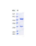
Anti-HDAC1 rabbit polyclonal antibody at 1:500 dilution Lane A: HepG2 Whole Cell Lysate Lane B: HeLa Whole Cell Lysate Lane C: HT-29 Whole Cell Lysate Lane D: COS7 Whole Cell Lysate Lysates/proteins at 30 μg per lane. Secondary Goat Anti-Rabbit IgG (H+L)/HRP at 1/10000 dilution. Developed using the ECL technique. Performed under reducing conditions. Predicted band size:55 kDa Observed band size:70 kDa
HDAC1 (Histone Deacetylase 1) is a Protein Coding gene. Histone Deacetylase 1 protein encoded by this gene belongs to the histone deacetylase/acuc/apha family and is a component of the histone deacetylase complex. It also interacts with retinoblastoma tumor-suppressor protein and this complex is a key element in the control of cell proliferation and differentiation. HDAC1 is a ubiquitous deacetylase enzyme responsible for the deacetylation of lysine residues on the N-terminal part of the core histones (H2A, H2B, H3, and H4). Together with metastasis-associated protein-2, it deacetylates p53 and modulates its effect on cell growth and apoptosis. HDAC1 is widely expressed in the lymph node, colon, and other tissues. Diseases associated with HDAC1 include Atrichia With Papular Lesions and Retinoblastoma.
Research Areas
Cancer Drug Targets
Related Pathways
Notch Signaling
p53 Pathway
Tags
Cancer Immunotherapy
Immune Checkpoint
Immunotherapy
Targeted Therapy
References
Beharry AW, Sandesara PB, Roberts BM, Ferreira LF, Senf SM, Judge AR. HDAC1 activates FoxO and is both sufficient and required for skeletal muscle atrophy. Journal of Cell Science. 2014;127(7):1441-1453.


-HD12OC3024-WB.png)