A synthetic peptide corresponding to the N-terminus of the Human Actin
Preparation
Produced in rabbits immunized with a synthetic peptide corresponding to the N-terminus of the Human Actin, and purified by antigen affinity chromatography.
Source
Polyclonal Rabbit IgG
Purification
Protein A & Antigen Affinity
Formulation
0.2 μm filtered solution in PBS
Conjugate
Unconjugated
Form
Liquid
Shipping
This antibody is shipped as liquid solution at ambient temperature. Upon receipt, store it immediately at the temperature recommended below.
Storage
This antibody can be stored at 2℃-8℃ for one month without detectable loss of activity. Antibody products are stable for twelve months from date of receipt when stored at -20℃ to -80℃. Preservative-Free. Avoid repeated freeze-thaw cycles.
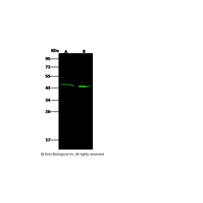
Anti-ACTA1 rabbit polyclonal antibody at 1:500 dilution Lane A: Hela Whole Cell Lysate Lane B: A549 Whole Cell Lysate Lysates/proteins at 30 μg per lane. Secondary Goat Anti-Rabbit IgG (H&L) /Dylight 800 at 1/10000 dilution. Developed using the Odyssey technique. Performed under reducing conditions. Predicted band size:42 kDa Observed band size:45 kDa
Anti-ACTA1 rabbit polyclonal antibody at 1:500 dilution Lane A: Hela Whole Cell Lysate Lane B: A549 Whole Cell Lysate Lysates/proteins at 30 μg per lane. Secondary Goat Anti-Rabbit IgG (H&L) /Dylight 800 at 1/10000 dilution. Developed using the Odyssey technique. Performed under reducing conditions. Predicted band size:42 kDa Observed band size:45 kDa
Anti-ACTA1 rabbit polyclonal antibody at 1:500 dilution Lane A: Hela Whole Cell Lysate Lane B: A549 Whole Cell Lysate Lysates/proteins at 30 μg per lane. Secondary Goat Anti-Rabbit IgG (H&L) /Dylight 800 at 1/10000 dilution. Developed using the Odyssey technique. Performed under reducing conditions. Predicted band size:42 kDa Observed band size:45 kDa
Anti-ACTA1 rabbit polyclonal antibody at 1:500 dilution Lane A: Hela Whole Cell Lysate Lane B: A549 Whole Cell Lysate Lysates/proteins at 30 μg per lane. Secondary Goat Anti-Rabbit IgG (H&L) /Dylight 800 at 1/10000 dilution. Developed using the Odyssey technique. Performed under reducing conditions. Predicted band size:42 kDa Observed band size:45 kDa
Anti-ACTA1 rabbit polyclonal antibody at 1:500 dilution Lane A: Hela Whole Cell Lysate Lane B: A549 Whole Cell Lysate Lysates/proteins at 30 μg per lane. Secondary Goat Anti-Rabbit IgG (H&L) /Dylight 800 at 1/10000 dilution. Developed using the Odyssey technique. Performed under reducing conditions. Predicted band size:42 kDa Observed band size:45 kDa
Nemaline myopathy (NM) is one of the most common congenital nondystrophic myopathies and is characterized by muscle weakness, often from birth. Mutations in ACTA1 are a frequent cause of NM (ie, NEM3). ACTA1 encodes alpha-actin 1, the main constituent of the sarcomeric thin filament. The mechanisms by which mutations in ACTA1 contribute to muscle weakness in NEM3 are incompletely understood. Mutations in skeletal muscle α-actin 1-encoding gene (ACTA1) cause autosomal dominant or recessive myopathies with marked clinical and pathological heterogeneity. Patients typically develop generalized or limb-girdle pattern of weakness, that clinically severe ACTA1-related myopathy can present with muscle morphological findings suggestive of cytoplasmic body myopathy in the absence of definite nemaline rods. The Asn94Lys mutation in skeletal muscle sarcomeric α-actin may be linked to this histological appearance.
title: Chlorogenic acid inhibits the proliferation of human lung cancer A549 cell lines by targeting annexin A2 in vitro and in vivo. authors: Lei Wang; Hongwu Du; Peng Chen journal: Biomedicine & pharmacotherapy = Biomedecine & pharmacotherapie date: 2021-02-18 pmid: 32882585 doi: 10.1016/j.biopha.2020.110673 cited_by_count: 80 techniques: Enzyme-linked Immunosorbent Assay; Imaging; Incubation; Membrane; SDS Page; Western Blot bioz_score: 92

-HD10MA2928-WB.png)
-HD10MA2928-1ug-prostate-IHC-P.png)
-HD10MA2928-2ug-skeletal-muscle-IHC-P.png)
-HD10MA2928-2ug-mouse-skeletal-muscle-IHC-P.png)
-HD10MA2928-2ug-rat-skeletal-muscle-IHC-P.png)