Human ALDH1A1 No cross-reactivity in ELISA with E.coli cell lysate
Immunogen
Recombinant Human ALDH1 protein (Catalog#11388-H07E)
Preparation
This antibody was produced from a hybridoma resulting from the fusion of a mouse myeloma with B cells obtained from a mouse immunized with purified, recombinant Human ALDH1 (rh ALDH1; Catalog#11388-H07E; NP_000680.2; Ser 2-Ser 501). The IgG fraction of the cell culture supernatant was purified by Protein A affinity chromatography .
Source
Monoclonal Mouse IgG1 Clone #5G9E6C9
Purification
Protein A
Formulation
0.2 μm filtered solution in PBS
Conjugate
Unconjugated
Form
Liquid
Shipping
This antibody is shipped as liquid solution at ambient temperature. Upon receipt, store it immediately at the temperature recommended below.
Storage
This antibody can be stored at 2℃-8℃ for one month without detectable loss of activity. Antibody products are stable for twelve months from date of receipt when stored at -20℃ to -80℃. Preservative-Free. Avoid repeated freeze-thaw cycles.
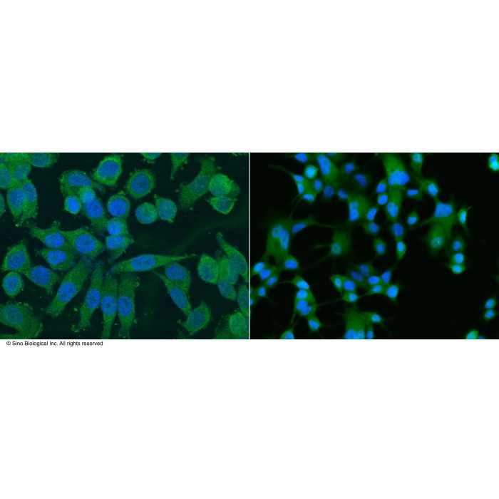
Immunofluorescence staining of Human ALDH1A1 in MCF7 or SKBR3 cells. Cells were fixed with 4% PFA, permeabilzed with 1% Triton X-100 in PBS, blocked with 10% serum, and incubated with mouse anti-Human ALDH1A1 monoclonal antibody (1:60). Then cells were stained with the Alexa Fluor® 488-conjugated Goat Anti-mouse IgG secondary antibody (left panel, captured by laser confocal scanning microscope; right panel, captured by fluorescence microscope), countstained with DAPI (blue). Positive staining was localized to cytoplasm.
Image 1 Alt Text
Human ALDH1A1 / ALDH1 Immunohistochemistry(IHC) 3013
Immunofluorescence staining of Human ALDH1A1 in MCF7 or SKBR3 cells. Cells were fixed with 4% PFA, permeabilzed with 1% Triton X-100 in PBS, blocked with 10% serum, and incubated with mouse anti-Human ALDH1A1 monoclonal antibody (1:60). Then cells were stained with the Alexa Fluor® 488-conjugated Goat Anti-mouse IgG secondary antibody (left panel, captured by laser confocal scanning microscope; right panel, captured by fluorescence microscope), countstained with DAPI (blue). Positive staining was localized to cytoplasm.
Immunofluorescence staining of Human ALDH1A1 in MCF7 or SKBR3 cells. Cells were fixed with 4% PFA, permeabilzed with 1% Triton X-100 in PBS, blocked with 10% serum, and incubated with mouse anti-Human ALDH1A1 monoclonal antibody (1:60). Then cells were stained with the Alexa Fluor® 488-conjugated Goat Anti-mouse IgG secondary antibody (left panel, captured by laser confocal scanning microscope; right panel, captured by fluorescence microscope), countstained with DAPI (blue). Positive staining was localized to cytoplasm.
Aldehyde dehydrogenase 1 family, member A1 (ALDH1A1), also known as Aldehyde dehydrogenase 1 (ALDH1), or Retinaldehyde Dehydrogenase 1 (RALDH1), is an enzyme that is expressed at high levels in stem cells and that has been suggested to regulate stem cell function. The retinaldehyde dehydrogenase (RALDH) subfamily of ALDHs, composed of ALDH1A1, ALDH1A2, ALDH1A3, and ALDH8A1, regulate development by catalyzing retinoic acid biosynthesis. The ALDH1A1 protein belongs to the aldehyde dehydrogenases family of proteins. Aldehyde dehydrogenase is the second enzyme of the major oxidative pathway of alcohol metabolism. ALDH1A1 also belongs to the group of corneal crystallins that help maintain the transparency of the cornea. Increased ALDH1A1 activity has been found in the stem cell populations of leukemia and some solid tumors. In tumor specimens, increased ALDH1A1 immunopositivity was found not only in secretory type cancer epithelial cells but also in neuroendocrine tumor populations. ALDH1 has been identified as a reliable marker of breast cancer stem cells. ALDH1 expression in primary cancer is an independent prognostic factor in node-positive breast cancer patients. ALDH1A1 plays a key role in normal hematopoiesis, and as a TLX1 transcriptional target, ALDH1A1 may contribute to the ability of this homeoprotein to alter cell fate and induce tumor growth.
References
Li T, et al. (2010). ALDH1A1 is a marker for malignant prostate stem cells and predictor of prostate cancer patients' outcome. Lab Invest. 90(2): 234-44.
Levi BP, et al. (2009) Aldehyde dehydrogenase 1a1 is dispensable for stem cell function in the mouse hematopoietic and nervous systems. Blood. 113(8): 1670-80.
Rahman FB, et al. (2006) Uncompetitive inhibition of Xenopus laevis aldehyde dehydrogenase 1A1 by divalent cations. Zoolog Sci. 23(3): 239-44.
Jester JV, et al. (1999). The cellular basis of corneal transparency: evidence for corneal crystallins. J Cell Sci. 112 ( Pt 5): 613-22.
title: A vascularized breast cancer spheroid platform for the ranked evaluation of tumor microenvironment-targeted drugs by light sheet fluorescence microscopy. authors: David Ascheid; Magdalena Baumann; ...; Erik Henke journal: Nature Communications date: 2024-04-27 pmid: 38678014 cited_by_count: 13 techniques: Clone Assay; FACS; Generated; Immunofluorescence; Plasmid Preparation; Staining; Transfection bioz_score: 93
title: A humanized breast cancer microorganoid platform for the ranked evaluation of tumor microenvironment-targeted drugs by light sheet fluorescence microscopy authors: Erik Henke; David Ascheid; ...; Katrin Heinze journal: Europe PMC date: 2023-12-02 doi: 10.21203/rs.3.rs-3627627 cited_by_count: 0 techniques: Clone Assay; FACS; Generated; Plasmid Preparation; Staining; Sterility; Transfection bioz_score: 93


-SKBR3-C(L).png)

