A synthetic peptide corresponding to the N-terminus of the Human APEX1 / AP / APEx / Ref-1
Preparation
Produced in rabbits immunized with a synthetic peptide corresponding to the N-terminus of the Human APEX1 / AP / APEx / Ref-1, and purified by antigen affinity chromatography.
Source
Polyclonal Rabbit IgG
Purification
Protein A & Antigen Affinity
Formulation
0.2 μm filtered solution in PBS
Conjugate
Unconjugated
Form
Liquid
Shipping
This antibody is shipped as liquid solution at ambient temperature. Upon receipt, store it immediately at the temperature recommended below.
Storage
This antibody can be stored at 2℃-8℃ for one month without detectable loss of activity. Antibody products are stable for twelve months from date of receipt when stored at -20℃ to -80℃. Preservative-Free. Avoid repeated freeze-thaw cycles.
Immunofluorescence staining of APEX1 in HeLa cells. Cells were fixed with 4% PFA,blocked with 10% serum, and incubated with rabbit anti-human APEX1 polyclonal antibody (1:1000) at 4℃ overnight. Then cells were stained with the Alexa Fluor®594-conjugated Goat Anti-rabbit IgG secondary antibody (green)Positive staining was localized to nucleus.
Image 1 Alt Text
Human APEX1/APE1/Ref-1 Immunofluorescence(IF) 3085
Immunofluorescence staining of APEX1 in HeLa cells. Cells were fixed with 4% PFA,blocked with 10% serum, and incubated with rabbit anti-human APEX1 polyclonal antibody (1:1000) at 4℃ overnight. Then cells were stained with the Alexa Fluor®594-conjugated Goat Anti-rabbit IgG secondary antibody (green)Positive staining was localized to nucleus.
Image 2 Alt Text
Human APEX1/APE1/Ref-1 Immunohistochemistry(IHC) 4696
Immunofluorescence staining of APEX1 in HeLa cells. Cells were fixed with 4% PFA,blocked with 10% serum, and incubated with rabbit anti-human APEX1 polyclonal antibody (1:1000) at 4℃ overnight. Then cells were stained with the Alexa Fluor®594-conjugated Goat Anti-rabbit IgG secondary antibody (green)Positive staining was localized to nucleus.
Image 3 Alt Text
Human APEX1/APE1/Ref-1 Immunohistochemistry(IHC) 4697
Immunofluorescence staining of APEX1 in HeLa cells. Cells were fixed with 4% PFA,blocked with 10% serum, and incubated with rabbit anti-human APEX1 polyclonal antibody (1:1000) at 4℃ overnight. Then cells were stained with the Alexa Fluor®594-conjugated Goat Anti-rabbit IgG secondary antibody (green)Positive staining was localized to nucleus.
Image 4 Alt Text
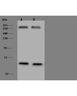
Human APEX1/APE1/Ref-1 Immunoprecipitation(IP) 5986
Immunofluorescence staining of APEX1 in HeLa cells. Cells were fixed with 4% PFA,blocked with 10% serum, and incubated with rabbit anti-human APEX1 polyclonal antibody (1:1000) at 4℃ overnight. Then cells were stained with the Alexa Fluor®594-conjugated Goat Anti-rabbit IgG secondary antibody (green)Positive staining was localized to nucleus.
APEX nuclease (multifunctional DNA repair enzyme) 1
Description
The enzyme is known to be a redox factor (Ref-1) stimulating DNA binding activity of AP-1 binding proteins such as Fos and Jun as well as a multifunctional DNA repair enzyme having 5' AP endonuclease, DNA 3' repair diesterase, 3'-5' exonuclease and DNA 3'-phosphatase activities.Although Apex mRNA was expressed ubiquitously, the levels varied significantly, suggesting organ- or tissue-specific expression of the Apex gene. The highest level was observed in the testis, relatively high levels in the thymus, spleen, kidney and brain, and the lowest level in the liver in rats. However, the present results suggested that APEX/Ref-1 gene product can interact with AP-1 binding proteins in brain, especially in the hippocampal formation, to regulate some brain functions by redox-activation.
References
Ono Y, et al . (1995) Developmental expression of APEX nuclease, a multifunctional DNA repair enzyme, in mouse brains. Brain Res Dev Brain Res.86 (1-2): 1-6.
Tan Y, et al . (1996) cDNA cloning of rat major AP endonuclease (APEX nuclease) and analyses of its mRNA expression in rat tissues. Acta Med Okayama. 50 (1): 53-60.
Yao M, et al . (1999) Genomic structure of the rat major AP endonuclease gene (Apex) with an adjacent putative O-sialoglycoprotease gene (Prsmg1/Gcpl1) and a processed Apex pseudogene (Apexp1). Acta Med Okayama. 53 (6): 245-52.


-HD09FE1505-1ug-HeLa-IF.png)
-HD09FE1505-0.5ug-placenta-IHC-P.png)
-HD09FE1505-0.5ug-breast-IHC-P.png)
-HD09FE1505-IP.png)
-HD09FE1505-WB.png)