Recombinant Human CALML5 protein (Catalog#11783-H20E)
Preparation
This antibody was obtained from a rabbit immunized with purified, recombinant Human CALML5 (rh CALML5; Catalog#11783-H20E; AAH39172.1; Met1-Glu146).
Source
Monoclonal Rabbit IgG Clone #119
Purification
Protein A
Formulation
0.2 μm filtered solution in PBS
Conjugate
Unconjugated
Form
Liquid
Shipping
This antibody is shipped as liquid solution at ambient temperature. Upon receipt, store it immediately at the temperature recommended below.
Storage
This antibody can be stored at 2℃-8℃ for one month without detectable loss of activity. Antibody products are stable for twelve months from date of receipt when stored at -20℃ to -80℃. Preservative-Free. Avoid repeated freeze-thaw cycles.
Synonyms: Anti-CLSP Antibody
Image / Experimental Information
Image 1 Description
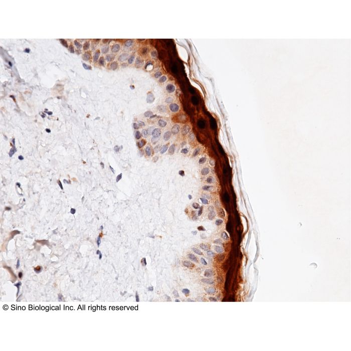
Immunochemical staining of human CALML5 in human skin with rabbit monoclonal antibody (1:200, formalin-fixed paraffin embedded sections). Positive staining was localized to epithelium.
Calmodulin-like protein 5, also known as Calmodulin-like skin protein, CALML5 and CLSP, is a protein which contains fourEF-hand domains. CALML5 / CLSP is particularly abundant in the epidermis where its expression is directly related to keratinocyte differentiation.The expression is very low in lung. CALML5 / CLSP binds calcium. It may be involved in terminal differentiation of keratinocytes. Coxsackievirus and adenovirus receptor (CAR) is a member of the immunoglobulin (Ig) superfamily and a component of epithelial tight junction. CAR functions as a primary receptor for coxsackievirus B and adenovirus (Ad) infection. CALML5 / CLSP is closely related to CAR. The structure and dynamics of human calmodulin-like skin protein CALML5 / CLSP have been characterized by NMR spectroscopy. The mobility of CALML5 / CLSP has been found to be different for the N-terminal and C-terminal domains. The N-terminal domain is characterized by four stable helices, which experience large fluctuations. This is shown to be due to mutations in the hydrophobic core. The overall N-terminal domain behavior is similar both in the full-length protein and in the isolated domain.
Related Pathways
Actin Dynamics Signaling Pathway
B Cell Receptor Signaling Pathway
T Cell Receptor Signaling Pathway
References
Mehul B., et al., 2000, J. Biol. Chem. 275:12841-12847.
Babini E., et al., 2006, Structure 14:1029-1038.
Kawabata,K. et al., 2007, Gene Ther. 14 (16):1199-207.