Recombinant Human ICAM3 / CD50 protein (Catalog#10333-H08H)
Preparation
This antibody was produced from a hybridoma resulting from the fusion of a mouse myeloma with B cells obtained from a mouse immunized with purified, recombinant Human CD50 / ICAM-3 (rh CD50 / ICAM-3; Catalog#10333-H08H; CAA49369.1; Met 1-His 485) and conjugated with APC under optimum conditions, the unreacted APC was removed.
Source
Monoclonal Mouse IgG2a Clone #02
Purification
Protein A
Formulation
PBS solution containing 0.5% BSA and 0.09% sodium azide
Concentration
5 μl/Test, 0.1 mg/ml
Conjugate
APC
Form
Liquid
Shipping
This antibody is shipped as liquid solution at ambient temperature. Upon receipt, store it immediately at the temperature recommended below.
Storage
This antibody can be stored at 2℃-8℃ for twelve months without detectable loss of activity. Protected from prolonged exposure to light. Do not freeze ! Sodium azide is toxic to cells and should be disposed of properly. Flush with large volumes of water during disposal.
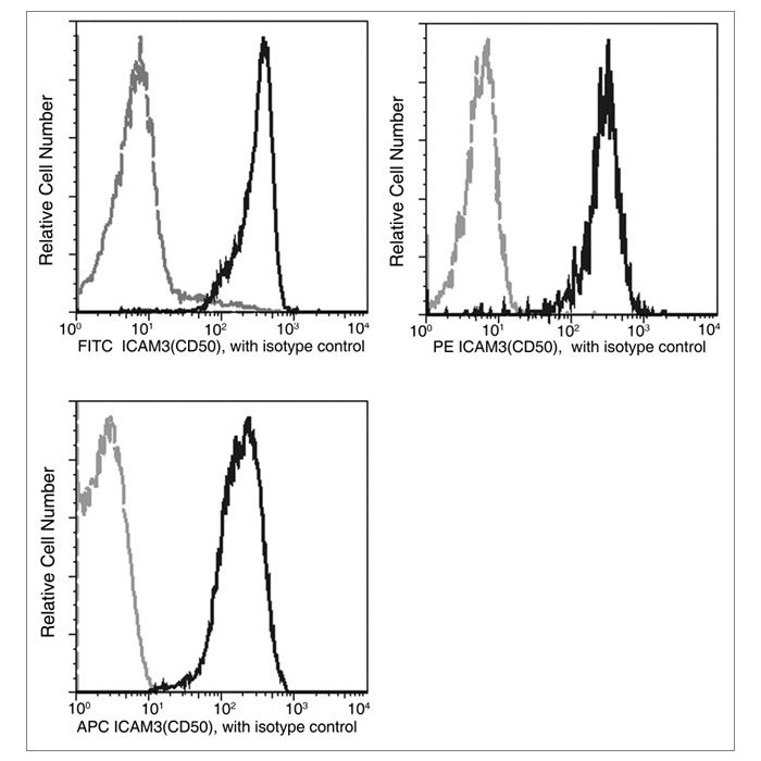
Profile of peripheral blood lymphocytes analyzed by flow cytometry. The fluorescence histograms were derived from events with the forward and side light-scatter characteristics of viable lymphocytes.
The protein ICAM-3, also known as CD50, is a member of the intercellular adhesion molecule (ICAM) family consisting of three members. It is a DC-SIGN ligand that is constitutively expressed on resting leukocytes and is thus an important molecule for the first immune response. ICAM-3 comprises five immunoglobulin-like domains and binds LFA-1 through its two N-terminal domains. It functions not only as an adhesion molecule but also as a potent signaling molecule. ICAM-3 binds to LFA-1 on antigen-presenting cells (APC) stabilizing the T cell-APC interaction, facilitating signaling through the CD3/TCR complex. However, recent evidence using cultured and transformed T cells suggests ICAM-3 may also function in signaling. It has been reported that the CD50 molecule can play a role in developing functionally mature T lymphocytes and its expression increases during the maturation process of T lymphocytes. Also, the interactions of ICAM-3 and LFA-1 facilitate HIV-1- induced virological synapse formation between T cells. ICAM-3 is associated with an increase in cellular radio-resistance and cancer cell proliferation. It could be considered as a candidate for anti-cancer drug development and as a cancer diagnostic marker.
References
Berney SM, et al. (1999) ICAM-3 (CD50) cross-linking augments signaling in CD3-activated peripheral human T lymphocytes. J Leukoc Biol. 65(6): 867-74.
van Buul JD, et al. (2004) ICAM-3 activation modulates cell-cell contacts of human bone marrow endothelial cells. J Vasc Res. 41(1): 28-37.
Sugino H. (2005) ICAM-3, a ligand for DC-SIGN, was duplicated from ICAM-1 in mammalian evolution, but was lost in the rodent genome. FEBS Lett. 579(13): 2901-6.
Park JK, et al. (2010) ICAM-3 enhances the migratory and invasive potential of human non-small cell lung cancer cells by inducing MMP-2 and MMP-9 via Akt and CREB. Int J Oncol. 36(1): 181-92.


-MM02-FITC-PE-APC-Lymphocytes.png)