A synthetic peptide corresponding to the center region of the human GFAP
Preparation
This antibody was obtained from a rabbit immunized with a synthetic peptide corresponding to the center region of the human GFAP.
Source
Monoclonal Rabbit IgG Clone #012
Purification
Protein A
Formulation
0.2 μm filtered solution in PBS
Conjugate
Unconjugated
Form
Liquid
Shipping
This antibody is shipped as liquid solution at ambient temperature. Upon receipt, store it immediately at the temperature recommended below.
Storage
This antibody can be stored at 2℃-8℃ for one month without detectable loss of activity. Antibody products are stable for twelve months from date of receipt when stored at -20℃ to -80℃. Preservative-Free. Avoid repeated freeze-thaw cycles.
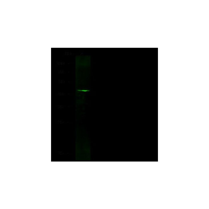
Anti-GFAP rabbit monoclonal antibody at 1:500 dilution Lane A: Mouse brain tissue lysate Lysates/proteins at 30 μg per lane. Secondary Goat Anti-Rabbit IgG H&L (Dylight800) at 1/10000 dilution. Developed using the Odyssey technique. Performed under reducing conditions. Predicted band size:50 kDa Observed band size:45 kDa
GFAP is a cell-specific marker which belongs to the intermediate filament family. It can distinguish astrocytes from other glial cells during development. GFAP is expressed in cells lacking fibronectin. It is a type III intermediate filaments protein which contains three domains: the head, rod and tail domains. GFAP functions in many important entral nervous system (CNS) processes, including cell communication and the functioning of the blood brain barrier. Improper GFAP regulation can cause multiple disorders. Defects in GFAP is related to Alexander disease which is a rare disorder of the central nervous system. It is a progressive leukoencephalopathy whose hallmark is the widespread accumulation of Rosenthal fibers which are cytoplasmic inclusions in astrocytes.
Research Areas
Neuroinflammation
References
Buniatian G, et al., 1998, Biology of the cell. 90(1): 53-61.
Chen YS, et al., 2011, Experimental Cell Research. 317(16): 2252-66.