WB (FAQ Protocol), ICC/IF (FAQ Protocol), IP (FAQ Protocol)
Species reactivity
Reacts with: Human
Specificity
Human Mouse, Rat (Species predicted to react based on 100% sequence homology)
Immunogen
A synthetic peptide corresponding to the N-terminus of the Human MEK1/MAP2K1/MKK1
Preparation
Produced in rabbits immunized with a synthetic peptide corresponding to the N-terminus of the Human MEK1/MAP2K1/MKK1, and purified by antigen affinity chromatography.
Source
Polyclonal Rabbit IgG
Purification
Protein A & Antigen Affinity
Formulation
0.2 μm filtered solution in PBS
Conjugate
Unconjugated
Form
Liquid
Shipping
This antibody is shipped as liquid solution at ambient temperature. Upon receipt, store it immediately at the temperature recommended below.
Storage
This antibody can be stored at 2℃-8℃ for one month without detectable loss of activity. Antibody products are stable for twelve months from date of receipt when stored at -20℃ to -80℃. Preservative-Free. Avoid repeated freeze-thaw cycles.
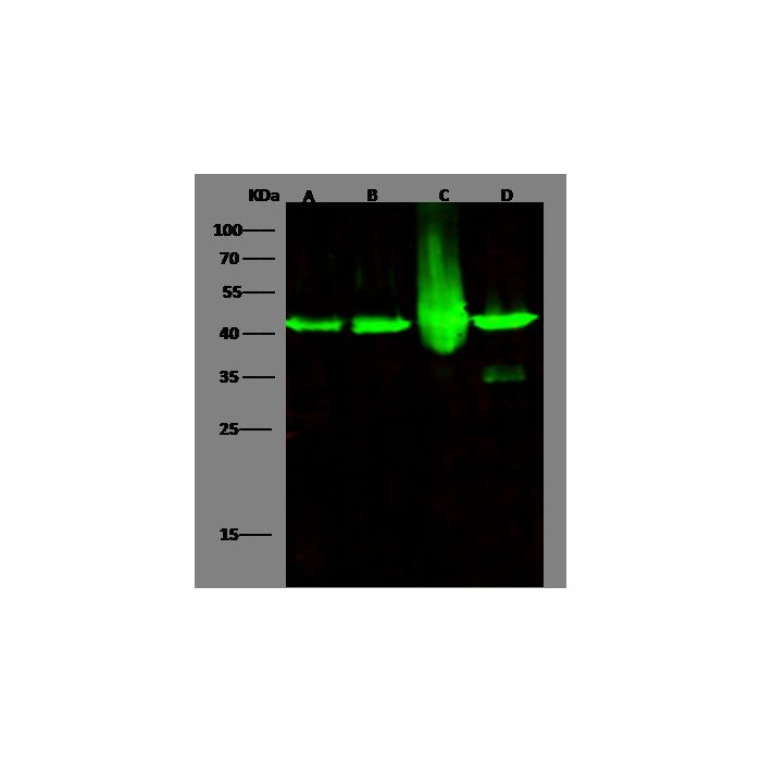
Anti-MAP2K1 rabbit polyclonal antibody at 1:500 dilution Lane A: HepG2 Whole Cell Lysate Lane B: Jurkat Whole Cell Lysate Lane C: Hela Whole Cell Lysate Lane D: A431 Whole Cell Lysate Lysates/proteins at 30 μg per lane. Secondary Goat Anti- Rabbit IgG H&L (Dylight 800) at 1/10000 dilution. Developed using the Odyssey technique. Performed under reducing conditions. Predicted band size:43 kDa Observed band size:43 kDa
Anti-MAP2K1 rabbit polyclonal antibody at 1:500 dilution Lane A: HepG2 Whole Cell Lysate Lane B: Jurkat Whole Cell Lysate Lane C: Hela Whole Cell Lysate Lane D: A431 Whole Cell Lysate Lysates/proteins at 30 μg per lane. Secondary Goat Anti- Rabbit IgG H&L (Dylight 800) at 1/10000 dilution. Developed using the Odyssey technique. Performed under reducing conditions. Predicted band size:43 kDa Observed band size:43 kDa
Image 2 Alt Text
Human MEK1/MAP2K1/MKK1 Immunofluorescence(IF) 17269
Anti-MAP2K1 rabbit polyclonal antibody at 1:500 dilution Lane A: HepG2 Whole Cell Lysate Lane B: Jurkat Whole Cell Lysate Lane C: Hela Whole Cell Lysate Lane D: A431 Whole Cell Lysate Lysates/proteins at 30 μg per lane. Secondary Goat Anti- Rabbit IgG H&L (Dylight 800) at 1/10000 dilution. Developed using the Odyssey technique. Performed under reducing conditions. Predicted band size:43 kDa Observed band size:43 kDa
Image 3 Alt Text
Human MEK1/MAP2K1/MKK1 Immunoprecipitation(IP) 17270
MEK1, also known as MAP2K1 and MKK1, is a member of the dual-specificity protein kinase family, which acts as a mitogen-activated protein (MAP) kinase kinase. MAP kinases, also known as extracellular signal-regulated kinases (ERKs), act as an integration point for multiple biochemical signals. MEK1 is widely expressed, with extremely low levels in the brain. It lies upstream of MAP kinases and stimulates the enzymatic activity of MAP kinases upon a wide variety of extra- and intracellular signals. As an essential component of the MAP kinase signal transduction pathway, MEK1 is involved in many cellular processes such as proliferation, differentiation, transcription regulation, and development. Binding extracellular ligands such as growth factors, cytokines, and hormones to their cell-surface receptors activates RAS and this initiates RAF1 activation. RAF1 then further activates the dual-specificity protein kinases MAP2K1 and MEK2. MEK1 has been shown to export PPARG from the nucleus. The MAPK cascade is also involved in the regulation of endosomal dynamics, including lysosome processing and endosome cycling through the perinuclear recycling compartment (PNRC), as well as in the fragmentation of the Golgi apparatus during mitosis. MKK1 catalyzes the concomitant phosphorylation of a threonine and a tyrosine residue in a Thr-Glu-Tyr sequence located in MAP kinases. Defects in MEK1 can cause Cardiofaciocutaneous Syndrome.
Research Areas
Cancer Drug Targets
Related Pathways
Death Receptor Signaling
EGFR Signaling Pathway
TGF-beta Signaling
VEGF Signaling Pathway
Actin Dynamics Signaling Pathway
B Cell Receptor Signaling Pathway
G Protein-coupled Receptors Signaling
Jak-Stat Signaling Pathway
MAPK-Erk Pathway
T Cell Receptor Signaling Pathway
Common G-Chain signaling pathway
IL1 signaling pathway
IL17 signaling pathway
Tags
Cancer Immunotherapy
Immune Checkpoint
Immunotherapy
Targeted Therapy
References
Rampoldi L, et al. (1998) Chromosomal localization of four MAPK signaling cascade genes: MEK1, MEK3, MEK4 and MEKK5. Cytogenet Cell Genet. 78(3-4):301-3.
Zheng CF, et al. (1993) Cloning and characterization of two distinct human extracellular signal-regulated kinase activator kinases, MEK1 and MEK2. J Biol Chem. 268(15):11435-9.
Nantel, et al. (1998) Interaction of the Grb10 adapter protein with the Raf1 and MEK1 kinases. J Biol Chem. 273(17):10475-84.
Hirata H, et al. (2012) I MicroRNA-1826 targets VEGFC, beta-catenin (CTNNB1) and MEK1 (MAP2K1) in human bladder cancer. Carcinogenesis. 33(1):41-8.


-HD09JL0103-WB.png)

