Mouse Rat (Species predicted to react based on 100% sequence homology)
Immunogen
A synthetic peptide corresponding to the C-terminus of the Mouse CAMKI
Preparation
Produced in rabbits immunized with a synthetic peptide corresponding to the C-terminus of the Mouse CAMKI, and purified by antigen affinity chromatography.
Source
Polyclonal Rabbit IgG
Purification
Protein A & Antigen Affinity
Formulation
0.2 μm filtered solution in PBS
Conjugate
Unconjugated
Form
Liquid
Shipping
This antibody is shipped as liquid solution at ambient temperature. Upon receipt, store it immediately at the temperature recommended below.
Storage
This antibody can be stored at 2℃-8℃ for one month without detectable loss of activity. Antibody products are stable for twelve months from date of receipt when stored at -20℃ to -80℃. Preservative-Free. Avoid repeated freeze-thaw cycles.
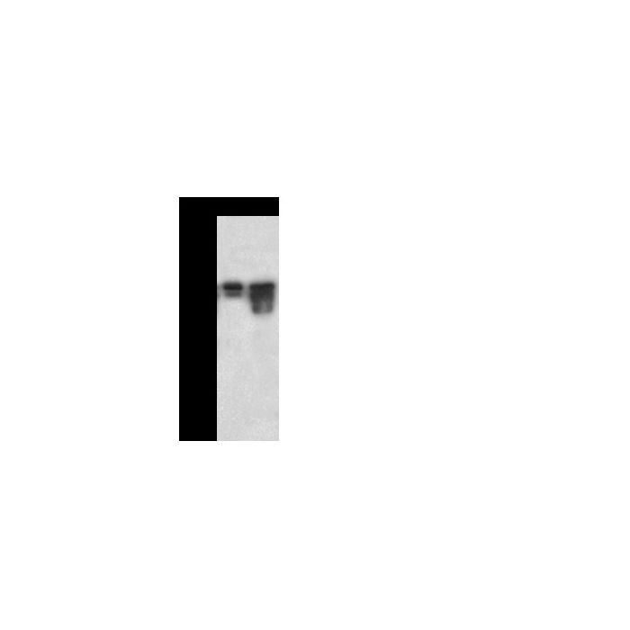
Anti-CAMK1 rabbit polyclonal antibody at 1:10000 dilution Lane A: HepG2 Whole Cell Lysate Lane B: Mouse brain tissue lysate Lysates/proteins at 30 μg per lane. Secondary Goat Anti-Rabbit IgG (H+L)/HRP at 1/10000 dilution. Developed using the ECL technique. Performed under reducing conditions. Predicted band size:41 kDa Observed band size:44 kDa
Calcium/calmodulin-dependent protein kinase or CaM kinases are serine/threonine-specific protein kinases that are primarily regulated by the Calcium/calmodulin complex. These kinases show a memory effect on activation. CaM kinases activity can outlast the intracellular calcium transient that is needed to activate it. Inneurons, this property is important for the induction of synaptic plasticity. Pharmacological inhibition of CaM kinases II blocks the induction of long-term potentiation. Upon activation, CaM kinases II phosphorylates postsynaptic glutamate receptors and changes the electrical properties of the synapse. Calcium/calmodulin-dependent protein kinase type 1D, also known as CaM kinase I delta, CaM kinase ID, CaMKI-like protein kinase, CKLiK and CAMK1D, is a member of theprotein kinase superfamily and CaMK subfamily. It contains oneprotein kinase domain. CAMK1D is broadly expressed. It is highly and mostly expressed in polymorphonuclear leukocytes (neutrophilic and eosinophilic granulocytes) while little or no expression is observed in monocytes and lymphocytes. Engineered overexpression of CAMK1D in non-tumorigenic breast epithelial cells led to increased cell proliferation, and molecular and phenotypic alterations indicative of epithelial-mesenchymal transition (EMT), including loss of cell-cell adhesions and increased cell migration and invasion. CAMK1D is a potential therapeutic target with particular relevance to clinically unfavorable basal-like tumors.
Research Areas
Calcium-binding Proteins and Related Molecules
Related Pathways
EGFR Signaling Pathway
B Cell Receptor Signaling Pathway
G Protein-coupled Receptors Signaling
References
Lisman, JE. et al., 1985, Proc Natl Acad Sci USA. 82 (9): 3055-7.
Bergamaschi, A. et al., 2008, Mol Oncol. 2 (4): 327-39.
White RB. et al., 2008, Physiological genomics, 33 (1): 41-9.
Schleinitz, D. et al., 2010, Horm Metab Res. 42 (1): 14-22.

-HD11DE1419-WB.png)