Human Mouse, Rat (Species predicted to react based on 100% sequence homology)
Immunogen
A synthetic peptide corresponding to the N-terminus of the Human Musashi-1
Preparation
Produced in rabbits immunized with a synthetic peptide corresponding to the N-terminus of the Human Musashi-1, and purified by antigen affinity chromatography.
Source
Polyclonal Rabbit IgG
Purification
Protein A & Antigen Affinity
Formulation
0.2 μm filtered solution in PBS
Conjugate
Unconjugated
Form
Liquid
Shipping
This antibody is shipped as liquid solution at ambient temperature. Upon receipt, store it immediately at the temperature recommended below.
Storage
This antibody can be stored at 2℃-8℃ for one month without detectable loss of activity. Antibody products are stable for twelve months from date of receipt when stored at -20℃ to -80℃. Preservative-Free. Avoid repeated freeze-thaw cycles.
Image / Experimental Information
Image 1 Description
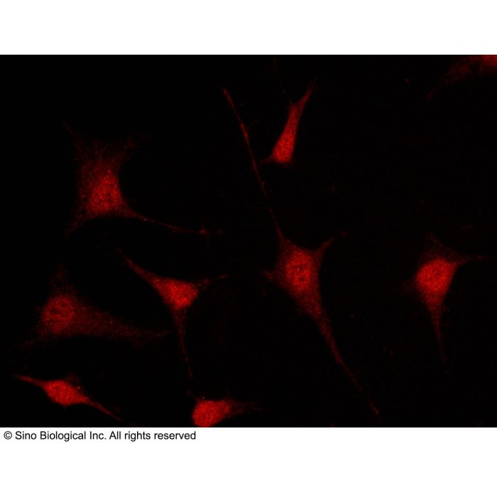
Immunofluorescence staining of MSI1 in Hela cells. Cells were fixed with 4% PFA, permeabilzed with 0.3% Triton X-100 in PBS,blocked with 10% serum, and incubated with rabbit anti-human MSI1 polyclonal antibody (1:1000) at 4℃ overnight. Then cells were stained with the Alexa Fluor®594-conjugated Goat Anti-rabbit IgG secondary antibody (red)Positive staining was localized to cytoplasm and nucleus.
Immunofluorescence staining of MSI1 in Hela cells. Cells were fixed with 4% PFA, permeabilzed with 0.3% Triton X-100 in PBS,blocked with 10% serum, and incubated with rabbit anti-human MSI1 polyclonal antibody (1:1000) at 4℃ overnight. Then cells were stained with the Alexa Fluor®594-conjugated Goat Anti-rabbit IgG secondary antibody (red)Positive staining was localized to cytoplasm and nucleus.
Immunofluorescence staining of MSI1 in Hela cells. Cells were fixed with 4% PFA, permeabilzed with 0.3% Triton X-100 in PBS,blocked with 10% serum, and incubated with rabbit anti-human MSI1 polyclonal antibody (1:1000) at 4℃ overnight. Then cells were stained with the Alexa Fluor®594-conjugated Goat Anti-rabbit IgG secondary antibody (red)Positive staining was localized to cytoplasm and nucleus.
Musashi RNA-binding protein 1 (MSI1) is highly expressed in certain cancer types and functions as a putative progenitor/stem cell marker. MSI1 belongs to a family of histone binding WD40-repeat proteins. MSI1 is a subunit of Polycomb group protein complexes and Chromatin assembly factor 1, and it interacts with the Retinoblastoma-related protein 1. Altered levels of MSI1 result in pleiotropic phenotypes, reflecting the complexity of MSI1 protein functions. MSI1 can bind to the chromatin of the drought-inducible downstream target RD20 and suggest a new role for MSI1 in the negative regulation of the Arabidopsis drought-stress response. The MSI1 (CAC3) gene of Saccharomyces cerevisiae has been implicated in diverse cellular functions, including suppression of the RAS/cAMP/protein kinase A signaling pathway, chromatin assembly and transcriptional co-repression.


-HB08NO0721-1ug-Hela-IF.png)
-HB08NO0721-2ug-hepatoma-IHC-P.png)
-HB08NO0721-2ug-brain-IHC-P.png)