Produced in rabbits immunized with E. coli-derived Human AARSD1 fragment, and purified by antigen affinity chromatography.
Source
Polyclonal Rabbit IgG
Purification
Protein A & Antigen Affinity
Formulation
PBS, pH7.0 with 0.03% Proclin300
Conjugate
Unconjugated
Form
Liquid
Shipping
This antibody is shipped as liquid solution at ambient temperature. Upon receipt, store it immediately at the temperature recommended below.
Storage
This antibody can be stored at 2℃-8℃ for one month without detectable loss of activity. Antibody products are stable for twelve months from date of receipt when stored at -20℃ to -80℃. Avoid repeated freeze-thaw cycles.
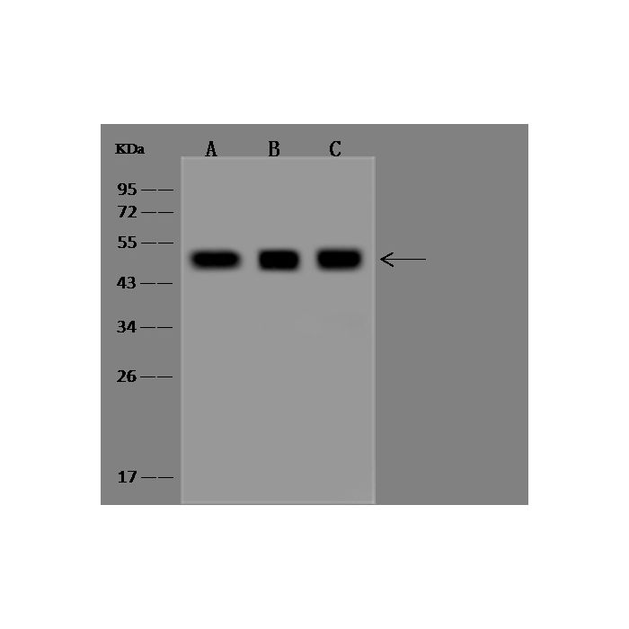
Anti-AARSD1 rabbit polyclonal antibody at 1:500 dilution Lane A: A549 Whole Cell Lysate Lane B: 293 Whole Cell Lysate Lane C: HepG2 Whole Cell Lysate Lysates/proteins at 30 μg per lane. Secondary Goat Anti-Rabbit IgG (H+L)/HRP at 1/10000 dilution. Developed using the ECL technique. Performed under reducing conditions. Predicted band size:45 kDa Observed band size:48 kDa
Anti-AARSD1 rabbit polyclonal antibody at 1:500 dilution Lane A: A549 Whole Cell Lysate Lane B: 293 Whole Cell Lysate Lane C: HepG2 Whole Cell Lysate Lysates/proteins at 30 μg per lane. Secondary Goat Anti-Rabbit IgG (H+L)/HRP at 1/10000 dilution. Developed using the ECL technique. Performed under reducing conditions. Predicted band size:45 kDa Observed band size:48 kDa
AARSD1 belongs to the class-II aminoacyl-tRNA synthetase family, Alax-L subfamily. AARSD1 binds 1 zinc ion per subunit functions in trans to edit the amino acid moiety from incorrectly charged tRNA(Ala). Four transcript variants have been described for AARSD1: NM_25267.3, NM_113642.2, NM_1142653.1 and NM_1142654.1. It has been determined that the latter two variants represent a distinct upstream locus, which is now represented by GeneID:1885848 (PTGES3L), while the former two variants represent readthrough transcripts between PTGES3L and this locus (AARSD1). The readthrough locus (PTGES3L-AARSD1) is now represented by GeneID:188585.
References
Tatham MH, et al. (2011) Comparative proteomic analysis identifies a role for SUMO in protein quality control. Oncogene. 19(17):2120-8.
Kim W, et al. (2011) Systematic and quantitative assessment of the ubiquitin-modified proteome. Mol Cell. 44(2):325-40.
Udeshi ND, et al. (2012) Methods for quantification of in vivo changes in protein ubiquitination following proteasome and deubiquitinase inhibition. Mol Cell Proteomics. 11(5):148-59.

-HD12SE2522-WB.png)
