Supplied as sterile 20mM Tris, 300mM NaCl, 10% Glycerol, pH 7.5. Please contact us for any concerns or special requirements. Please refer to the specific buffer information in the hardcopy of datasheet or the lot-specific COA.
Endotoxin
< 1.0 EU per μg protein as determined by the LAL method.
Shipping
Liquid. It is shipped out with blue ice.
Stability & Storage
Samples are stable for up to twelve months from date of receipt at -20℃ to -80℃. Store it under sterile conditions at -20℃ to -80℃. It is recommended that the protein be aliquoted for optimal storage. Avoid repeated freeze-thaw cycles.
Reconstitution
To find reconstitution info, click on 'COA' at the top of the page, enter the lot number and product size for the lot-specific COA.
Purity
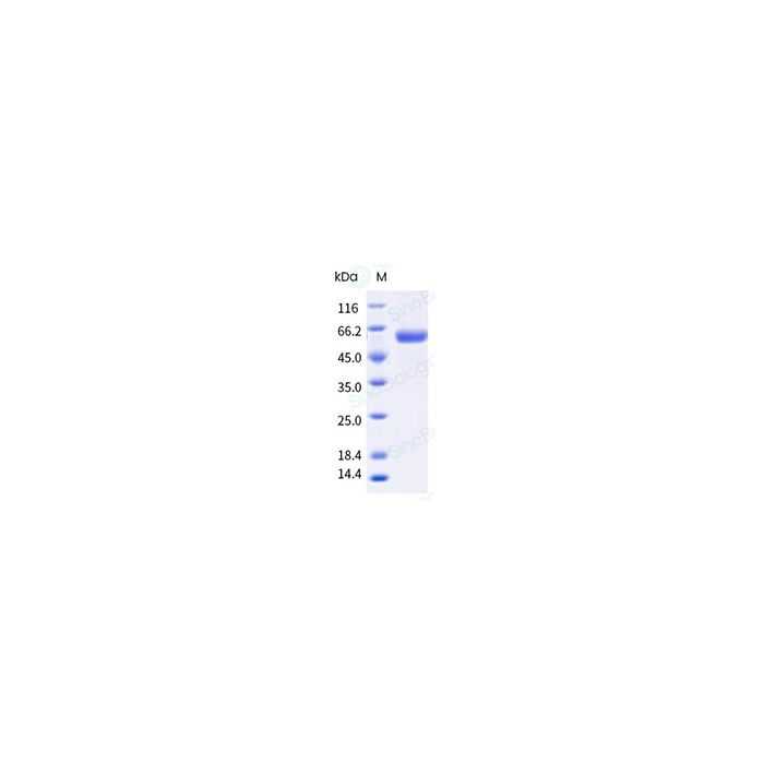
≥ 90 % as determined by SDS-PAGE.
Activity
Measured by its ability to cleave a fluorogenic substrate, 2'-(4-Methylumbelliferyl)-α-D-N-acetylneuraminic acid. The specific activity is >4000pmols/min/ug
Protein Construction
A DNA sequence encoding the Influenza A virus (A/Hong Kong/01/1968(H3N2)) Neuraminidase / NA (EPI901060) (Val30-Ile469) was expressed with a vasodilator-stimulated phosphoprotein tetramerization domain at the N-terminus and a polyhistidine tag at the C-terminus.
Accession#
EPI901060
Expressed Host
Baculovirus-Insect Cells
Species
H3N2
Predicted N Terminal
Asp
Molecule Mass
The recombinant NA subunit of the Influenza A virus (A/Hong Kong/01/1968(H3N2)) consists 486 amino acids and predicts a molecular mass of 54.82 kDa. It migrates as an approximately 58.9 kDa band in SDS-PAGE under reducing conditions.
Neuraminidases are enzymes that cleave sialic acid groups from glycoproteins. Influenza neuraminidase is a type of neuraminidase found on the surface of influenza viruses that enables the virus to be released from the host cell. Influenza neuraminidase is composed of four identical subunits arranged in a square. It is normally attached to the virus surface through a long protein stalk. The active sites are in a deep depression on the upper surface. They bind to polysaccharide chains and clip off the sugars at the end. The surface of neuraminidase is decorated with several polysaccharide chains that are similar to the polysaccharide chains that decorate our cell surface proteins. Neuraminidase (NA) and hemagglutinin (HA) are major membrane glycoproteins found on the surface of the influenza virus. Hemagglutinin binds to the sialic acid-containing receptors on the surface of host cells during initial infection and at the end of an infectious cycle. Neuraminidase, on the other hand, cleaves the HA-sialic acid bondage from the newly formed virions and the host cell receptors during budding. Neuraminidase thus is described as a receptor-destroying enzyme that facilitates virus release and efficient spread of the progeny virus from cell to cell. Influenza antibody and influenza antibodies are very important research tools for influenza diagnosis, influenza vaccine development, and anti-influenza virus therapy development. The monoclonal or polyclonal antibody can be raised with protein based antigen or peptide-based antigen. Antibodies raised with protein-based antigen could have better specificity and/or binding affinity than antibodies raised with peptide based antigen, but the cost associated with the recombinant protein antigen is usually higher. Anti-influenza virus hemagglutinin (HA) monoclonal antibody or polyclonal antibody can be used for ELISA assay, western blotting detection, Immunohistochemistry (IHC), flow cytometry, neutralization assay, hemagglutinin inhibition assay, and early diagnosis of influenza viral infection. Sino Biological has developed state-of-the-art monoclonal antibody development technology platforms: mouse monoclonal antibody and rabbit monoclonal antibody. Our rabbit monoclonal antibody platform is one of a kind and offers some unique advantages over mouse monoclonal antibodies, such as high affinity, low cross-reactivity with rabbit polyclonal antibodies.
References
Sardet C., et al. ,(1989), Molecular cloning, primary structure, and expression of the human growth factor-activatable Na+/H+ antiporter. Cell 56:271-280.
Sardet C., et al. , (1990), Growth factors induce phosphorylation of the Na+/H+ antiporter, glycoprotein of 110 kD.Science 247:723-726.
Tse C.-M., et al. ,(1991), Molecular cloning and expression of a cDNA encoding the rabbit ileal villus cell basolateral membrane Na+/H+ exchanger.EMBO J. 10:1957-1967.