Recombinant Human / Mouse HIST1H3A Protein (Catalog#11231-HNAE)
Preparation
Produced in rabbits immunized with purified, recombinant Human / Mouse HIST1H3A (rh HIST1H3A; Catalog#11231-HNAE; NP_003520.1; Met1-Ala136). HIST1H3A specific IgG was purified by Human / Mouse HIST1H3A affinity chromatography.
Source
Polyclonal Rabbit IgG
Purification
Protein A & Antigen Affinity
Formulation
0.2 μm filtered solution in PBS
Conjugate
Unconjugated
Form
Liquid
Shipping
This antibody is shipped as liquid solution at ambient temperature. Upon receipt, store it immediately at the temperature recommended below.
Storage
This antibody can be stored at 2℃-8℃ for one month without detectable loss of activity. Antibody products are stable for twelve months from date of receipt when stored at -20℃ to -80℃. Preservative-Free. Avoid repeated freeze-thaw cycles.
Image / Experimental Information
Image 1 Description
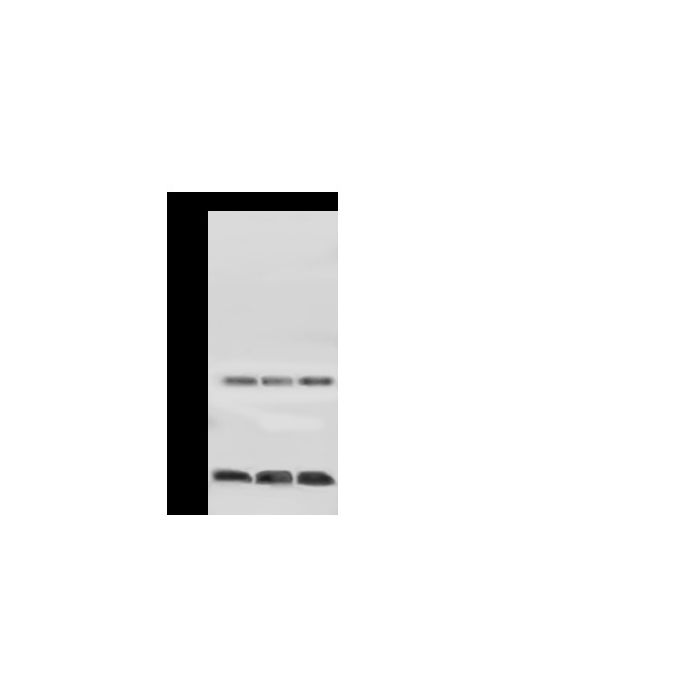
Anti-H3A rabbit polyclonal antibody at 1:500 dilution Lane A: HeLa Whole Cell Lysate Lane B: NIH-3T3 Whole Cell Lysate Lane C: Jurkat Whole Cell Lysate Lysates/proteins at 30 μg per lane. Secondary Goat Anti-Rabbit IgG (H+L)/HRP at 1/10000 dilution. Developed using the ECL technique. Performed under reducing conditions. Predicted band size:15 kDa Observed band size:15 kDa
Anti-H3A rabbit polyclonal antibody at 1:500 dilution Lane A: HeLa Whole Cell Lysate Lane B: NIH-3T3 Whole Cell Lysate Lane C: Jurkat Whole Cell Lysate Lysates/proteins at 30 μg per lane. Secondary Goat Anti-Rabbit IgG (H+L)/HRP at 1/10000 dilution. Developed using the ECL technique. Performed under reducing conditions. Predicted band size:15 kDa Observed band size:15 kDa
Anti-H3A rabbit polyclonal antibody at 1:500 dilution Lane A: HeLa Whole Cell Lysate Lane B: NIH-3T3 Whole Cell Lysate Lane C: Jurkat Whole Cell Lysate Lysates/proteins at 30 μg per lane. Secondary Goat Anti-Rabbit IgG (H+L)/HRP at 1/10000 dilution. Developed using the ECL technique. Performed under reducing conditions. Predicted band size:15 kDa Observed band size:15 kDa
Anti-H3A rabbit polyclonal antibody at 1:500 dilution Lane A: HeLa Whole Cell Lysate Lane B: NIH-3T3 Whole Cell Lysate Lane C: Jurkat Whole Cell Lysate Lysates/proteins at 30 μg per lane. Secondary Goat Anti-Rabbit IgG (H+L)/HRP at 1/10000 dilution. Developed using the ECL technique. Performed under reducing conditions. Predicted band size:15 kDa Observed band size:15 kDa
Anti-H3A rabbit polyclonal antibody at 1:500 dilution Lane A: HeLa Whole Cell Lysate Lane B: NIH-3T3 Whole Cell Lysate Lane C: Jurkat Whole Cell Lysate Lysates/proteins at 30 μg per lane. Secondary Goat Anti-Rabbit IgG (H+L)/HRP at 1/10000 dilution. Developed using the ECL technique. Performed under reducing conditions. Predicted band size:15 kDa Observed band size:15 kDa
Histone H3.1, also known as HIST1H3A, HIST1H3B, HIST1H3C, HIST1H3D, HIST1H3E, HIST1H3F, HIST1H3G, HIST1H3H, HIST1H3I, HIST1H3J, is a member of the histone H3 family which is a core component of nucleosome. It is expressed during the S phase, then expression strongly decreases as cell division slows down during the process of differentiation. Nucleosomes wrap and compact DNA into chromatin, limiting DNA accessibility to the cellular machinery which requires DNA as a template. Histones thereby play a central role in transcription regulation, DNA repair, DNA replication, and chromosomal stability. DNA accessibility is regulated via a complex set of post-translational modifications of histones, also called histone code, and nucleosome remodeling. Histones are basic nuclear proteins that are responsible for the nucleosome structure of the chromosomal fiber in eukaryotes. This structure consists of approximately 146 bp of DNA wrapped around an octamer composed of pairs of each of the four core histones (H2A, H2B, H3, and H4). The chromatin fiber is further compacted through the interaction of a linker histone, H1, with the DNA between the nucleosomes to form higher-order chromatin structures.
Related Pathways
MAPK-Erk Pathway
P38 MAPK Signaling Pathway
References
Lachner M, et al., 2001, Nature 410 (6824): 116-20.
Koessler H, et al., 2003, DNA Cell Biol. 22 (4): 233-41.
Macdonald N. et al., 2005, Mol. Cell 20: 199-211.
Hyllus D. et al., 2007, Genes Dev. 21: 3369-3380.
Garcia BA. et al., 2007, J. Biol. Chem. 282:7641-7655.
Yu L.-R. et al., 2007, J. Proteome Res. 6: 4150-4162.

-HD12JA1117-WB.png)
-HD12JA1117-1000-human-breast-carcinoma-IHC-P.png)
-HD12JA1117-1000-human-colon-carcinoma-IHC-P.png)
-HD12JA1117-1000-HeLa-IF.png)
