Recombinant Human Beta-2 microglobulin/B2M Protein (Catalog#11976-H08H)
Preparation
This antibody was produced from a hybridoma resulting from the fusion of a mouse myeloma with B cells obtained from a mouse immunized with purified, recombinant Human Beta-2 microglobulin/B2M (rh Beta-2 microglobulin/B2M; Catalog#11976-H08H; NP_004039.1; Met1-Met119). The IgG fraction of the cell culture supernatant was purified by Protein A affinity chromatography.
Source
Monoclonal Mouse IgG1 Clone #34
Purification
Protein A
Formulation
0.2 μm filtered solution in PBS with 10% Trehalose, pH7.0
Conjugate
Unconjugated
Form
Liquid
Shipping
This antibody is shipped as liquid solution at ambient temperature. Upon receipt, store it immediately at the temperature recommended below.
Storage
This antibody can be stored at 2℃-8℃ for one month without detectable loss of activity. Antibody products are stable for twelve months from date of receipt when stored at -20℃ to -80℃. Preservative-Free. Avoid repeated freeze-thaw cycles.
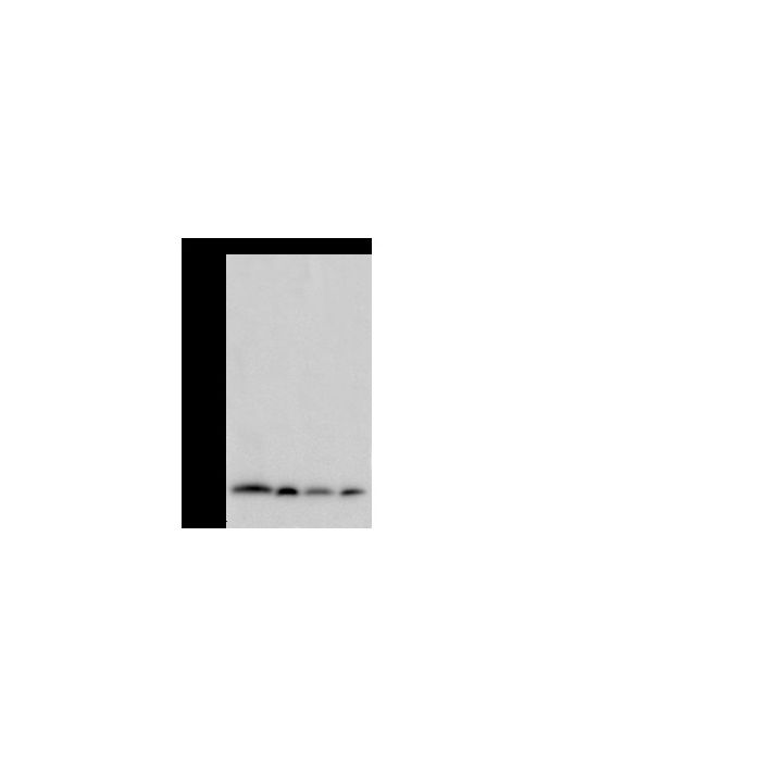
Anti-B2M mouse monoclonal antibody at 1:500 dilution Lane A: A431 Whole Cell Lysate Lane B: U937 Whole Cell Lysate Lane C: Raji Whole Cell Lysate Lane D: HeLa Whole Cell Lysate Lysates/proteins at 30 μg per lane. Secondary Goat Anti-Mouse IgG (H+L)/HRP at 1/10000 dilution. Developed using the ECL technique. Performed under reducing conditions. Predicted band size:14 kDa Observed band size:14 kDa
Image 1 Alt Text
Human Beta-2 microglobulin Western blot (WB) 20067
Anti-B2M mouse monoclonal antibody at 1:500 dilution Lane A: A431 Whole Cell Lysate Lane B: U937 Whole Cell Lysate Lane C: Raji Whole Cell Lysate Lane D: HeLa Whole Cell Lysate Lysates/proteins at 30 μg per lane. Secondary Goat Anti-Mouse IgG (H+L)/HRP at 1/10000 dilution. Developed using the ECL technique. Performed under reducing conditions. Predicted band size:14 kDa Observed band size:14 kDa
Image 2 Alt Text
Human Beta-2 microglobulin Immunohistochemistry(IHC) 20068
Anti-B2M mouse monoclonal antibody at 1:500 dilution Lane A: A431 Whole Cell Lysate Lane B: U937 Whole Cell Lysate Lane C: Raji Whole Cell Lysate Lane D: HeLa Whole Cell Lysate Lysates/proteins at 30 μg per lane. Secondary Goat Anti-Mouse IgG (H+L)/HRP at 1/10000 dilution. Developed using the ECL technique. Performed under reducing conditions. Predicted band size:14 kDa Observed band size:14 kDa
Image 3 Alt Text
Human Beta-2 microglobulin Immunohistochemistry(IHC) 20069
Anti-B2M mouse monoclonal antibody at 1:500 dilution Lane A: A431 Whole Cell Lysate Lane B: U937 Whole Cell Lysate Lane C: Raji Whole Cell Lysate Lane D: HeLa Whole Cell Lysate Lysates/proteins at 30 μg per lane. Secondary Goat Anti-Mouse IgG (H+L)/HRP at 1/10000 dilution. Developed using the ECL technique. Performed under reducing conditions. Predicted band size:14 kDa Observed band size:14 kDa
Image 4 Alt Text
Human Beta-2 microglobulin Immunofluorescence(IF) 20070
Anti-B2M mouse monoclonal antibody at 1:500 dilution Lane A: A431 Whole Cell Lysate Lane B: U937 Whole Cell Lysate Lane C: Raji Whole Cell Lysate Lane D: HeLa Whole Cell Lysate Lysates/proteins at 30 μg per lane. Secondary Goat Anti-Mouse IgG (H+L)/HRP at 1/10000 dilution. Developed using the ECL technique. Performed under reducing conditions. Predicted band size:14 kDa Observed band size:14 kDa
Image 5 Alt Text
Human Beta-2 microglobulin Flow Cytometry (FC) 21285
B2M, also known as β2-Microglobulin or CDABP0092, is a component of MHC class I molecules found expression in all nucleated cells (excludes red blood cells). The major function of MHC class I moleculesis is to display fragments of proteins from within the cell to T-cells and cells containing foreign proteins will be attacked. B2M (β2-Microglobulin) is a low molecular weight protein. It was demonstrated that B2M (β2-Microglobulin) was localized in the membranes of nucleated cells and was found to be associated with HL-A antigens. B2M (β2- Microglobulin) is present in free form in various body fluids and as a subunit of histocompatibility antigens on cell surfaces lateral to the α3 chain. Unlike α3, β2 has no transmembrane region. Directly above β2 lies the α1 chain, which itself is lateral to the α2. In the absence of B2M (β2 microglobulin), very limited amounts of MHC class I (classical and non-classical) molecules can be detected on the surface. In the absence of MHC class I, CD8 T cells, a subset of T cells involved in the development of acquired immunity cannot develop. Low levels of B2M (β2 microglobulin) can indicate non-progression of HIV.
References
Poulik MD, et al. (1979) Beta 2-Microglobulin: methods and clinical applications. CRC Ctit Rev Clin Lab Sci. 10(3): 225-45.
Poulik MD, et al. (1975) Beta2-Microglobulins. Contemp Top Mol Immunol. 4: 157-204.
Berggard I. (1976) Beta2-Microglobulins: isolation, properties, and distribution. Fed Proc. 35(5): 1167-70.




-MM34-Purified-Lymphocytes.png)