A synthetic peptide corresponding to the N-terminus of the human beta-tubulin
Preparation
It is a chimeric monoclonal antibody combining the constant domains of the rabbit IgG molecule with mouse variable regions. The variable region was obtained from a mouse immunized with a synthetic peptide corresponding to the N-terminus of the human beta-tubulin. The antibody was produced using recombinant antibody technology.
Source
Monoclonal Rabbit IgG Clone #05
Purification
Protein A
Formulation
0.2 μm filtered solution in PBS
Conjugate
Unconjugated
Form
Liquid
Shipping
This antibody is shipped as liquid solution at ambient temperature. Upon receipt, store it immediately at the temperature recommended below.
Storage
This antibody can be stored at 2℃-8℃ for one month without detectable loss of activity. Antibody products are stable for twelve months from date of receipt when stored at -20℃ to -80℃. Preservative-Free. Avoid repeated freeze-thaw cycles.
Image / Experimental Information
Image 1 Description
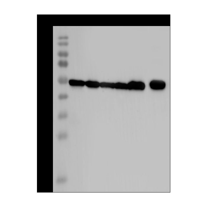
Anti-Beta-Tubulin rabbit monoclonal antibody at 1:30000 dilution Lane A: Jurkat Whole Cell Lysate Lane B: HeLa Whole Cell Lysate Lane C: HepG2 Whole Cell Lysate Lane D: A549 Whole Cell Lysate Lane E: Mouse brain tissue lysate Lane F: Rat brain tissue lysate Lysates/proteins at 30 μg per lane. Secondary Goat Anti-Rabbit IgG (H+L)/HRP at 1/10000 dilution. Developed using the ECL technique. Performed under reducing conditions. Predicted band size:50 kDa Observed band size:54 kDa
Anti-Beta-Tubulin rabbit monoclonal antibody at 1:30000 dilution Lane A: Jurkat Whole Cell Lysate Lane B: HeLa Whole Cell Lysate Lane C: HepG2 Whole Cell Lysate Lane D: A549 Whole Cell Lysate Lane E: Mouse brain tissue lysate Lane F: Rat brain tissue lysate Lysates/proteins at 30 μg per lane. Secondary Goat Anti-Rabbit IgG (H+L)/HRP at 1/10000 dilution. Developed using the ECL technique. Performed under reducing conditions. Predicted band size:50 kDa Observed band size:54 kDa
Image 2 Alt Text
Human Beta-Tubulin Immunohistochemistry(IHC) 42452
Anti-Beta-Tubulin rabbit monoclonal antibody at 1:30000 dilution Lane A: Jurkat Whole Cell Lysate Lane B: HeLa Whole Cell Lysate Lane C: HepG2 Whole Cell Lysate Lane D: A549 Whole Cell Lysate Lane E: Mouse brain tissue lysate Lane F: Rat brain tissue lysate Lysates/proteins at 30 μg per lane. Secondary Goat Anti-Rabbit IgG (H+L)/HRP at 1/10000 dilution. Developed using the ECL technique. Performed under reducing conditions. Predicted band size:50 kDa Observed band size:54 kDa
Image 3 Alt Text
Human Beta-Tubulin Immunohistochemistry(IHC) 42453
Anti-Beta-Tubulin rabbit monoclonal antibody at 1:30000 dilution Lane A: Jurkat Whole Cell Lysate Lane B: HeLa Whole Cell Lysate Lane C: HepG2 Whole Cell Lysate Lane D: A549 Whole Cell Lysate Lane E: Mouse brain tissue lysate Lane F: Rat brain tissue lysate Lysates/proteins at 30 μg per lane. Secondary Goat Anti-Rabbit IgG (H+L)/HRP at 1/10000 dilution. Developed using the ECL technique. Performed under reducing conditions. Predicted band size:50 kDa Observed band size:54 kDa
Image 4 Alt Text
Human Beta-Tubulin Immunohistochemistry(IHC) 42454
Anti-Beta-Tubulin rabbit monoclonal antibody at 1:30000 dilution Lane A: Jurkat Whole Cell Lysate Lane B: HeLa Whole Cell Lysate Lane C: HepG2 Whole Cell Lysate Lane D: A549 Whole Cell Lysate Lane E: Mouse brain tissue lysate Lane F: Rat brain tissue lysate Lysates/proteins at 30 μg per lane. Secondary Goat Anti-Rabbit IgG (H+L)/HRP at 1/10000 dilution. Developed using the ECL technique. Performed under reducing conditions. Predicted band size:50 kDa Observed band size:54 kDa
Beta-Tubulin is a subunit of tubulin. Tubulin is one of several members of a small family of globular proteins. It is the major constituent of microtubules. There are two most common members of the tubulin family: alpha-tubulin and beta-tubulin, and together their dimers form microtubules. The dimers of alpha- and beta-tubulin bind to GTP and assemble onto the (+) ends of microtubules while in the GTP-bound state. After the dimer is incorporated into the microtubule, the molecule of GTP bound to the beta -tubulin subunit eventually hydrolyzes into GDP through inter-dimer contacts along the microtubule protofilament. Beta-tubulin faces the plus end of the microtubule while alpha-tubulin faces the minus end. Dimers bound to GTP tend to assemble into microtubules, while dimers bound to GDP tend to fall apart. Loading controls are usually proteins that exhibit high-level, constitutive expression in the cell type or sample you are examining. This ensures constant expression levels. Thus “housekeeping genes” are frequently chosen for use as loading controls. It is also important that the protein chosen as a loading control has a different molecular weight than the protein of interest so that the bands are distinct and expression levels quantifiable. Popular loading control detection antibodies include anti-β-Actin monoclonal or polyclonal antibodies, anti-COX-4, anti-GAPDH, anti-Tubulin and anti-VDAC/Porin antibodies.
References
Williams RC Jr. et al., 1999. Anal Biochem. 275(2): 265-7.
Nogales E. et al., 1998. Nat Struct Biol. 5(6): 451-8.
title: Both E6 and E7 in HPV16 Promote the Glucose Uptake of GLUT1 in Lung Cancer Cells By Repressing NDRG2 Expression and Further Nuclear Translocation of β-Catenin authors: Xin Wang; Ming-Zhe Wu; ...; Guang-Ping Wu journal: Europe PMC date: 2021-06-08 doi: 10.21203/rs.3.rs-573879 cited_by_count: 0 techniques: Electrochemiluminescence; Imaging; Incubation; Transfection; Western Blot bioz_score: 90
title: MAGE-A11 is activated through TFCP2/ZEB1 binding sites de-methylation as well as histone modification and facilitates ESCC tumor growth authors: Shina Liu; Fei Liu; ...; Meixiang Sang journal: Oncotarget date: 2018-01-09 pmid: 29423052 doi: 10.18632/oncotarget.22973 pmcid: 5790469 cited_by_count: 10 techniques: Chromatin Immunoprecipitation; Western Blot bioz_score: 90

-HA18JU1102-WB.png)


