WB (FAQ Protocol), ELISA (FAQ Protocol), IP (FAQ Protocol)
Species reactivity
Reacts with: Human
Specificity
Human MAX
Immunogen
Recombinant Human MYC associated factor X protein (Catalog#12885-H20B)
Preparation
Produced in rabbits immunized with purified, recombinant Human MYC associated factor X (rh MYC associated factor X; Catalog#12885-H20B; NP_002373; Met1-Ser160). MYC associated factor X specific IgG was purified by Human MYC associated factor X affinity chromatography.
Source
Polyclonal Rabbit IgG
Purification
Protein A & Antigen Affinity
Formulation
0.2 μm filtered solution in PBS
Conjugate
Unconjugated
Form
Liquid
Shipping
This antibody is shipped as liquid solution at ambient temperature. Upon receipt, store it immediately at the temperature recommended below.
Storage
This antibody can be stored at 2℃-8℃ for one month without detectable loss of activity. Antibody products are stable for twelve months from date of receipt when stored at -20℃ to -80℃. Preservative-Free. Avoid repeated freeze-thaw cycles.
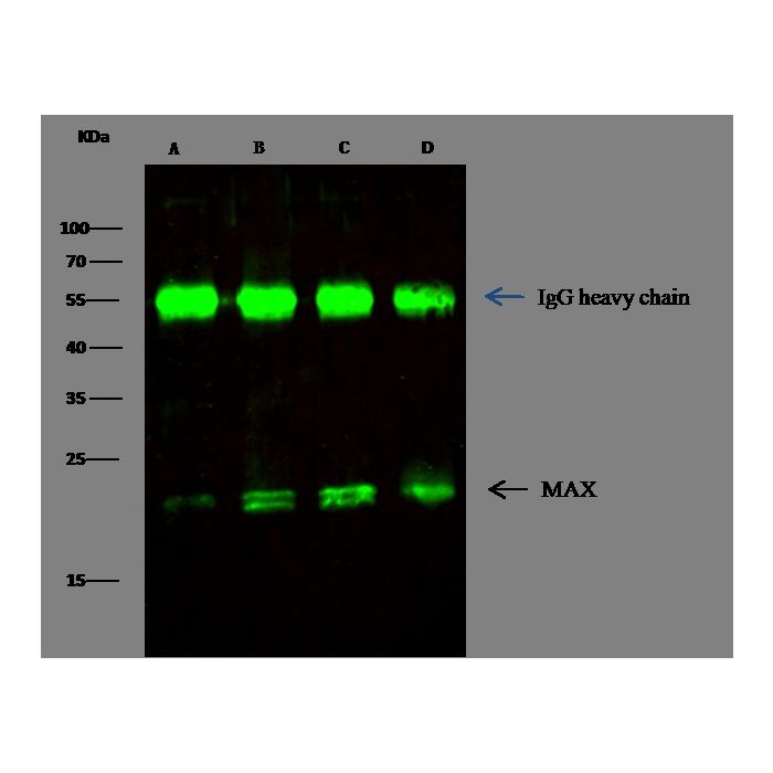
MAX was immunoprecipitated using: Lane A:0.5 mg Jurkat Whole Cell Lysate Lane B:0.5 mg Hela Whole Cell Lysate Lane C:0.5 mg 293T Whole Cell Lysate Lane D:0.5 mg A549 Whole Cell Lysate 2 µL anti-MAX rabbit polyclonal antibody and 15 μl of 50 % Protein G agarose. Primary antibody: Anti-MAX rabbit polyclonal antibody,at 1:330 dilution Secondary antibody: Dylight 800-labeled antibody to rabbit IgG (H+L), at 1:5000 dilution Developed using the odssey technique. Performed under reducing conditions. Predicted band size: 23 kDa Observed band size: 23 kDa
Image 1 Alt Text
Human MYC associated factor X Immunoprecipitation(IP) 6044
MAX was immunoprecipitated using: Lane A:0.5 mg Jurkat Whole Cell Lysate Lane B:0.5 mg Hela Whole Cell Lysate Lane C:0.5 mg 293T Whole Cell Lysate Lane D:0.5 mg A549 Whole Cell Lysate 2 µL anti-MAX rabbit polyclonal antibody and 15 μl of 50 % Protein G agarose. Primary antibody: Anti-MAX rabbit polyclonal antibody,at 1:330 dilution Secondary antibody: Dylight 800-labeled antibody to rabbit IgG (H+L), at 1:5000 dilution Developed using the odssey technique. Performed under reducing conditions. Predicted band size: 23 kDa Observed band size: 23 kDa
Image 2 Alt Text
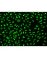
Human MYC associated factor X Western blot (WB) 6670
MYC associated factor X contains 1 basic helix-loop-helix (bHLH) domain and belongs to the MAX family. It is highly expressed in the brain, heart, and lung while lower levels are seen in the liver, kidney, and skeletal muscle. MYC associated factor X can form homodimers and heterodimers with other family members, which include Mad, Mxi1, and Myc. Myc is an oncoprotein implicated in cell proliferation, differentiation, and apoptosis. The homodimers and heterodimers compete for a common DNA target site (the E box) and rearrangement among these dimer forms provides a complex system of transcriptional regulation. MYC associated factor X may also repress transcription via the recruitment of a chromatin remodeling complex containing H3 'Lys-9' histone methyltransferase activity. Multiple alternatively spliced transcript variants have been described for MYC associated factor X gene but the full-length nature for some of them is unknown.
Research Areas
Apoptosis Transcription Factors and Regulators
Related Pathways
P38 MAPK Signaling Pathway
References
Mac Partlin M, et al. (2003) Interactions of the DNA mismatch repair proteins MLH1 and MSH2 with c-MYC and MAX. Oncogene. 22(6):819-25.
Cheng SW, et al. (1999) c-MYC interacts with INI1/hSNF5 and requires the SWI/SNF complex for transactivation function. Nat enet. 22(1):102-5.
McMahon SB, et al. (1998) The novel ATM-related protein TRRAP is an essential cofactor for the c-Myc and E2F oncoproteins. Cell. 94(3):363-74.

-HB07JA1109-IP.png)
-HB07JA1109-WB.png)