Recombinant Human N-Cadherin / CD325 / CDH2 protein (Catalog#11039-H08H)
Preparation
This antibody was obtained from a rabbit immunized with purified, recombinant Human N-Cadherin / CD325 / CDH2 (rh N-Cadherin / CD325 / CDH2; Catalog#11039-H08H; NP_001783.2; Met1-Ala724) and conjugated with APC under optimum conditions, the unreacted APC was removed.
Source
Monoclonal Rabbit IgG Clone #020
Purification
Protein A
Formulation
PBS solution containing 0.5% BSA and 0.09% sodium azide
Concentration
10 μl/Test, 0.1 mg/ml
Conjugate
APC
Form
Liquid
Shipping
This antibody is shipped as liquid solution at ambient temperature. Upon receipt, store it immediately at the temperature recommended below.
Storage
This antibody can be stored at 2℃-8℃ for twelve months without detectable loss of activity. Protected from prolonged exposure to light. Do not freeze ! Sodium azide is toxic to cells and should be disposed of properly. Flush with large volumes of water during disposal.
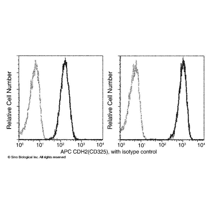
Flow cytometry analysis of N-Cadherin on HeLa cells. HeLa cells were harvested with (Left) or without (Right) trypsinization (please note, the epitope is sensitive to trypsin) and stained with either Rabbit IgG isotype control FITC (dashed line) or Rabbit Anti-Human CD325 antibody APC (solid line) at matching concentrations.
Cadherins are calcium-dependent cell adhesion proteins, and they preferentially interact with themselves in a homophilic manner in connecting cells. Cadherin 2 (CDH2), also known as N-Cadherin (neuronal) (NCAD), is a single-pass transmembrane protein and a cadherin containing 5 cadherin domains. N-Cadherin displays a ubiquitous expression pattern but with different expression levels between endocrine cell types. CDH2 (NCAD) has been shown to play an essential role in normal neuronal development, which is implicated in an array of processes including neuronal differentiation and migration, and axon growth and fasciculation. In addition, N-Cadherin expression was upregulated in human HSC during activation in culture, and function or expression blocking of N-Cadherin promoted apoptosis. During apoptosis, N-Cadherin was cleaved into 20-100 kDa fragments. It may provide a novel target for therapies that are directed toward intimal proliferative disorders, including restenosis and vascular bypass graft failure. N-Cadherin is associated with tumor aggressiveness and metastatic potential and may contribute to tumor progression.
Research Areas
Early Mesodermal Lineage Markers
Related Pathways
Canonical Wnt Pathway
References
Jones M, et al. (2002) N-cadherin upregulation and function in response of smooth muscle cells to arterial injury. Arterioscler Thromb Vasc Biol. 22(12): 1972-7.
Nagi C, et al. (2005) N-cadherin expression in breast cancer: correlation with an aggressive histologic variant--invasive micropapillary carcinoma. Breast Cancer Res Treat. 94(3): 225-35.
Schrick C, et al. (2007) N-cadherin regulates cytoskeletally associated IQGAP1/ERK signaling and memory formation. Neuron. 55(5): 786-98.
Li K, et al. (2010) Downregulation of N-cadherin expression inhibits invasiveness, arrests cell cycle and induces cell apoptosis in esophageal squamous cell carcinoma. Cancer Invest. 28(5): 479-86.



-R020-APC-HeLa.png)