Produced in rabbits immunized with E. coli-derived Human MDH1 fragment, and purified by antigen affinity chromatography.
Source
Polyclonal Rabbit IgG
Purification
Protein A & Antigen Affinity
Formulation
PBS, pH7.0 with 0.03% Proclin300
Conjugate
Unconjugated
Form
Liquid
Shipping
This antibody is shipped as liquid solution at ambient temperature. Upon receipt, store it immediately at the temperature recommended below.
Storage
This antibody can be stored at 2℃-8℃ for one month without detectable loss of activity. Antibody products are stable for twelve months from date of receipt when stored at -20℃ to -80℃. Avoid repeated freeze-thaw cycles.
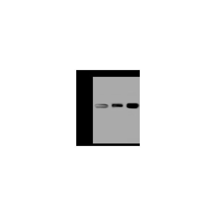
Anti-MDH1 rabbit polyclonal antibody at 1:500 dilution Lane A: Jurkat Whole Cell Lysate Lane B: HeLa Whole Cell Lysate Lane C: HepG2 Whole Cell Lysate Lysates/proteins at 30 μg per lane. Secondary Goat Anti-Rabbit IgG (H+L)/HRP at 1/10000 dilution. Developed using the ECL technique. Performed under reducing conditions. Predicted band size:36 kDa Observed band size:36 kDa
Anti-MDH1 rabbit polyclonal antibody at 1:500 dilution Lane A: Jurkat Whole Cell Lysate Lane B: HeLa Whole Cell Lysate Lane C: HepG2 Whole Cell Lysate Lysates/proteins at 30 μg per lane. Secondary Goat Anti-Rabbit IgG (H+L)/HRP at 1/10000 dilution. Developed using the ECL technique. Performed under reducing conditions. Predicted band size:36 kDa Observed band size:36 kDa
Malate dehydrogenases 1(MDH1 / MDHA) is a soluble form of malate dehydrogenases. Malate dehydrogenases (MDH) is a group of multimeric enzymes consisting of identical subunits usually organized as either dimer or tetramers with subunit molecular weights of 30-35 kDa. MDH has been isolated from different sources including archaea, eubacteria, fungi, plants, and mammals. MDH catalyzes the NAD/NADH-dependent interconversion of the substrates malate and oxaloacetate. This reaction plays a key part in the malate/aspartate shuttle across the mitochondrial membrane, and in the tricarboxylic acid cycle within the mitochondrial matrix. The enzymes share a common catalytic mechanism and their kinetic properties are similar, which demonstrates a high degree of structural similarity. The three-dimensional structures and elements essential for catalysis are conserved between mitochondrial and cytoplasmic forms of MDH in eukaryotic cells even though these isoenzymes are only marginally related at the level of the primary structure.
References
Minarik P, et al . (2002) Malate dehydrogenases--structure and function. Gen Physiol Biophys. 21 (3): 257-65.
Musrati RA, et al . (1998) Malate dehydrogenase: distribution, function and properties. Gen Physiol Biophys. 17 (3): 193-210.
Hall MD, et al . (1992) Crystal structure of Escherichia coli malate dehydrogenase. A complex of the apoenzyme and citrate at 1.87 A resolution. J Mol Biol. 226 (3): 867-82.


-HD14JL1338-WB.png)
