Recombinant Mouse ACO2 / Aconitase 2 protein (Catalog#51015-M20B)
Preparation
Produced in rabbits immunized with purified, recombinant Mouse ACO2 / Aconitase 2 (Catalog#51015-M20B; Q99KI0; Gln28-Gln780). ACO2 / Aconitase 2 specific IgG was purified by Mouse ACO2 / Aconitase 2 affinity chromatography.
Source
Polyclonal Rabbit IgG
Purification
Protein A & Antigen Affinity
Formulation
0.2 μm filtered solution in PBS
Conjugate
Unconjugated
Form
Liquid
Shipping
This antibody is shipped as liquid solution at ambient temperature. Upon receipt, store it immediately at the temperature recommended below.
Storage
This antibody can be stored at 2℃-8℃ for one month without detectable loss of activity. Antibody products are stable for twelve months from date of receipt when stored at -20℃ to -80℃. Preservative-Free. Avoid repeated freeze-thaw cycles.
A homozygous missense mutation was identified in the ACO2 gene (c.1240T>G p.Phe414Val) that segregated with HSP complicated by intellectual disability and microcephaly. Lymphoblastoid cell lines of homozygous carrier patients revealed significantly decreased activity of the mitochondrial aconitase enzyme and defective mitochondrial respiration. ACO2 encodes mitochondrial aconitase, an essential enzyme in the Krebs cycle. Recessive mutations in this gene have been previously associated with cerebellar ataxia. We found homozygous or compound heterozygous missense and frameshift mutations in the gene encoding mitochondrial aconitase (ACO2), a tricarboxylic acid cycle enzyme, catalysing interconversion of citrate into isocitrate. Unlike wild type ACO2, all mutant ACO2 proteins failed to complement the respiratory growth of a yeast aco1-deletion strain. The study shows that autosomal recessive ACO2 mutations can cause either isolated or syndromic optic neuropathy. This observation identifies ACO2 as the second gene responsible for non-syndromic autosomal recessive optic neuropathies and provides evidence for a genetic overlap between isolated and syndromic forms, giving further support to the view that optic atrophy is a hallmark of defective mitochondrial energy supply.
References
Robbins AH, et al. (1989) The structure of aconitase. Proteins. 5 (4): 289-312.
Lauble H, et al. (1992) Crystal structures of aconitase with isocitrate and nitroisocitrate bound. Biochemistry. 31 (10): 2735-48.
Robbins AH, et al. (1989) Structure of activated aconitase: formation of the 4Fe-4S cluster in the crystal. Proc Natl Acad Sci. 86 (10): 3639-43.

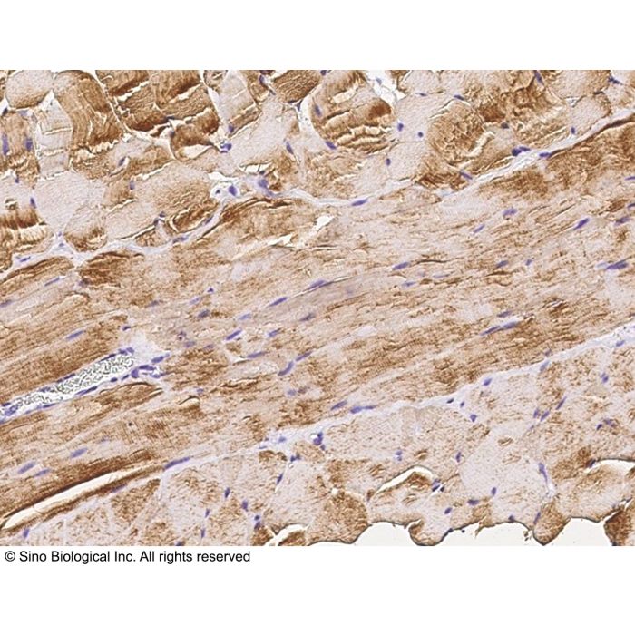



-HB08AU0825-0.2ug-mouse-skeletal-muscle-IHC-P.png)
-HB08AU0825-0.2ug-mouse-kidney-IHC-P.png)
-HB08AU0825-0.2ug-mouse-heart-IHC-P.png)
-pAb(A)-HB08AU0825-WB.png)
-Pab(A)-HB08AU0825-IP.png)