A synthetic peptide corresponding to the center region of the Human HLA-DR
Preparation
Produced in rabbits immunized with a synthetic peptide corresponding to the center region of the Human HLA-DR, and purified by antigen affinity chromatography.
Source
Polyclonal Rabbit IgG
Purification
Protein A & Antigen Affinity
Formulation
0.2 μm filtered solution in PBS
Conjugate
Unconjugated
Form
Liquid
Shipping
This antibody is shipped as liquid solution at ambient temperature. Upon receipt, store it immediately at the temperature recommended below.
Storage
This antibody can be stored at 2℃-8℃ for one month without detectable loss of activity. Antibody products are stable for twelve months from date of receipt when stored at -20℃ to -80℃. Preservative-Free. Avoid repeated freeze-thaw cycles.
Image / Experimental Information
Image 1 Description
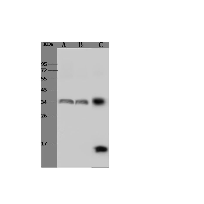
Anti-HLA-DR rabbit polyclonal antibody at 1:500 dilution Lane A: Raji Whole Cell Lysate Lane B: Daudi Whole Cell Lysate Lane C: HuT-78 Whole Cell Lysate Lysates/proteins at 30 μg per lane. Secondary Goat Anti-Rabbit IgG (H+L)/HRP at 1/10000 dilution. Developed using the ECL technique. Performed under reducing conditions. Predicted band size:29 kDa Observed band size:33 kDa
Anti-HLA-DR rabbit polyclonal antibody at 1:500 dilution Lane A: Raji Whole Cell Lysate Lane B: Daudi Whole Cell Lysate Lane C: HuT-78 Whole Cell Lysate Lysates/proteins at 30 μg per lane. Secondary Goat Anti-Rabbit IgG (H+L)/HRP at 1/10000 dilution. Developed using the ECL technique. Performed under reducing conditions. Predicted band size:29 kDa Observed band size:33 kDa
Anti-HLA-DR rabbit polyclonal antibody at 1:500 dilution Lane A: Raji Whole Cell Lysate Lane B: Daudi Whole Cell Lysate Lane C: HuT-78 Whole Cell Lysate Lysates/proteins at 30 μg per lane. Secondary Goat Anti-Rabbit IgG (H+L)/HRP at 1/10000 dilution. Developed using the ECL technique. Performed under reducing conditions. Predicted band size:29 kDa Observed band size:33 kDa
Anti-HLA-DR rabbit polyclonal antibody at 1:500 dilution Lane A: Raji Whole Cell Lysate Lane B: Daudi Whole Cell Lysate Lane C: HuT-78 Whole Cell Lysate Lysates/proteins at 30 μg per lane. Secondary Goat Anti-Rabbit IgG (H+L)/HRP at 1/10000 dilution. Developed using the ECL technique. Performed under reducing conditions. Predicted band size:29 kDa Observed band size:33 kDa
Anti-HLA-DR rabbit polyclonal antibody at 1:500 dilution Lane A: Raji Whole Cell Lysate Lane B: Daudi Whole Cell Lysate Lane C: HuT-78 Whole Cell Lysate Lysates/proteins at 30 μg per lane. Secondary Goat Anti-Rabbit IgG (H+L)/HRP at 1/10000 dilution. Developed using the ECL technique. Performed under reducing conditions. Predicted band size:29 kDa Observed band size:33 kDa
HLA-DR is a major histocompatibility complex (MHC) class II antigen presentation molecule, critical for the activation of lymphocytes and the orchestration of adaptive immune responses. HLA-DR is normally expressed on antigen-presenting cells including monocytes, macrophages, dendritic cells and B cells, but expression can be induced on epithelial cells and tumour cells in response to inflammatory conditions. MHC class II antigen presentation molecules are responsible for presentation of exogenous antigens to CD4 helper T cells. HLA-DR expression in tumours has been positively associated with enhanced lymphocytic infiltration. HLA-DR has been used as a surrogate marker of immune competence in various clinical studies, and its loss in circulation has been linked with susceptibility to post-surgical infection and sepsis.
References
Dunne MR, et al. (2020) Characterising the prognostic potential of hla-dr during colorectal cancer development. Cancer Immunol Immunother 69 (8): 1577-1588.