General Information
| Intended Use | The kit applies for the detection of mouse IFN-γ at the cellular level by counting IFN-γ-secreting cells. |
| PRINCIPLE OF THE ASSAY | The enzyme-linked immunospot (ELISPOT) assay employs a sandwich ELISA-based methodology. A 96-well polyvinylidene difluoride (PVDF) membrane plate, pre-coated with mouse IFN-γ-specific monoclonal antibodies, captures mouse IFN-γ secreted by activated cells. Following cell removal and washing, biotin-conjugated secondary antibodies targeting IFN-γ are added to form antibody-antigen-antibody "sandwich" complexes. Subsequent incubation with streptavidin-alkaline phosphatase (ALP) conjugates enables biotin-mediated binding. Addition of chromogenic substrate culminates in the formation of insoluble spots at the membrane interface, with each spot representing an individual IFN-γ-secreting cell. Spot quantification is achieved by automated ELISPOT analysis or manual counting using a microscope. |
| KIT CONTENTS AND STORAGE | Please use the kit before the expiration date. Components Amount Preparation instructions storage Pre-coated microplate 1plate (96 tests) Once removed from vacuum bag, the plate should be used as soon as possible. Vacuum storage can store at 2-8℃ until expiration date. Detection Antibody 1 vial Dilute the detection antibody 1:350 in PBS containing 0.5% fetal bovine serum (PBS-0.5% FBS) before use. Dilute fresh as needed. Primary liquid are stable at 2 - 8℃ until expiration date. To be reconstituted, the working fluid is used within the working day and discard. So dilute fresh as needed. Streptavidin-ALP 1 vial Dilute the Streptavidin-ALP 1:170 in PBS-0.5% FBS. BCIP/NBT-plus Dilution 1 bottle BCIP/NBT-plus Solution A and the Dilution should be mixed together in a 1:250 volume, followed by the addition of Solution B in the same volume as Solution Aand mixed gently.The volume ratio of Solution A:Dilution:Solution B is 1:250:1. For example: pipette 40 µl of Solution A into 10 ml of the Dilution buffer, mix thoroughly, then add 40 µl of Solution B and mix gently. Dilute fresh as needed. BCIP/NBT-plus Solution A 1 bottle BCIP/NBT-plus Solution B 1 bottle |
| ASSAY PROCEDURE | 1. Plate Set-up (sterile conditions) Remove the plate from the sealed package and wash 4 times with sterile PBS (200 µl/well). Condition the plate with sterile RPMI-1640 culture media (200 µl/well) containing 10% fetal bovine serum (FBS) asused for the cell suspensions, Incubate for at least 30 minutes at room temperature. 2. Incubation of cells in plate (sterile conditions) Empty the plate, then add stimulus followed by the 100 µl cell suspensions to each well. The concentration of cell and stimulus should be adjusted according to the actual situation.The recommended group settings are as follows. a. Positive control: Fewer cells such as 1×10 5 cells/wellare needed for polyclonal activators, e.g., ConA (0.25-2.5 µg/ml). b. Negative control: The cell concentration is kept consistent with the experimental wells in the absence of stimulus. c. Background control: No cells, no positive stimulus, add to the medium. d. Experimental group: The recommended cell number per well is 1.5–2.5 × 10 5 , and add your own stimulus as required by the experiment (prepare the stimulus using culture medium). Note: It is recommended to setduplicate wells for samples to be tested. Put the plate in a 37℃ and 5% CO 2 incubator for 18-48 hours.Do not move the plate during this period and it is recommended to wrap the plate in aluminum foil to avoid evaporation. 3. Detection of spots Note: Use PBS containing 0.5% FBS (PBS-0.5% FBS) for dilution ofthe detection antibody andStreptavidin-ALP. The PBS should be filtered (0.2 μm) for optimal results.Avoid the inclusion of Tween or other detergents in the washing and incubation buffers. Remove the cells by emptying the plate and wash 5 times with PBS, 200 µl/well. Note: Soak in the PBS for 1 minute to ensure thorough cleaning. The same requirement applies to all subsequent washing steps. Dilute the detection antibody 1:350 in PBS-0.5% FBS. Add 100 µl/well and incubate for 2 hours at room temperature. Note: Diluted detection antibody can be filtered (0.2 µm) to reduce the risk of unspecific background. Empty the plate and wash 5 times with PBS, 200 µl/well. Dilute the Streptavidin-ALP 1:170 in PBS-0.5% FBS and add 100 µl/well. Incubate for 1hour at room temperature. Empty the plate and wash 5 times with PBS, 200 µl/well. Filter the ready-to-use substrate solution (BCIP/NBT-plus) through a 0.45 µm filter andadd 100 µl/well. Develop until distinct spots emerge in positive wells (usually 5-30 minutes). Note: Too long the color development time will cause the background color in the experimental well to become darker. Stop color development by washing extensively in tap water. If desirable, remove theunderdrain (the soft plastic under the plate) and rinse the underside of the membrane. Leave the plate to dry. Plates should be completely dry before analysis. Inspect and count spots in an ELISpot reader or in a microscope. Note: Do not dry the microplate at a temperature higher than 37°C; this may cause cracking of the membrane filters. For the option to re-analyze later, store plate in the dark at room temperature. |
| TROUBLESHOOTING GUIDE | Problem Possible reason Solution Dark background of the PVDF membrane *Plate is insufficiently washed * The membrane is wet *Increase the washing times to ensure soaking time *Microplates should be analyzeduntil the PVDF membranes are completely dry Spots appear in the negative control wells *Endotoxins or other contaminants in the medium, serum, or DMSO may activate immune cells *The cells are contaminated *Cell viability is low *Use endotoxin-free reagents *Pay attention to aseptic procedures in the step of Plate Set-up and Incubation of cells in plate *Ensure high viability (recommended viability > 90%) as well as functionality of the cell sample Spots are irregular in shape and severely clumped *Cell disruption *Cell aggregated *Optimize the cell separation process to ensure cell activity *The cell suspension is thoroughly mixed toensure a unicellular state Positive Control spots were low and fuzzy *Enzyme activity is decreased *The temperature of the Substrate reagent is low and oxidized *The titer of the stimulus is low *Increase enzyme concentration *Balance the color reagent to room temperature before use, do not use reagents that have oxidized precipitation *Increase the concentration of stimulus The spots are unevenly positioned and have an edge effect *Shake the cell plate after plating, causing cell displacement *Add the stimulus too vigorously to flush the cell suspension *Stack the plates in the incubator *After plating, try not to shake the plate, and gently pan it into the incubator *Use the method of adding the stimulus first and then the cells *Place each plate individually on the shelf to allow an even distribution of heat to each microwell Not all spots are counted in the crowded well despite algorithm and parameter adjustments *Spots are confluent. Single spots cannot be discriminated *Decrease the number of cells added per well |
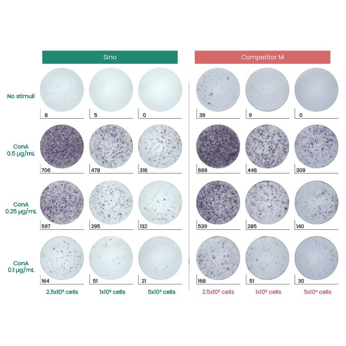
| Performance | Sino Competitor M The statistical histogram and representative plate images of mIFN-γ ELISpot assays. Splenocytes isolated from BALB/c mice were plated in graded densities (5×10 4 –2.5×10 5 cells/well) and stimulated with Concanavalin A (ConA) for 20 h. Wells containing cells without stimulation were designated as the negative control. |
Synonyms: If2f; Ifg; IFN-g; Ifng
Image / Experimental Information
| Image 1 Alt Text | EK50709-IFN-G-Sino-Mabtech |
| Image 1 URL | Source image |
Background Information
| Full Name | interferon, gamma |
| Description | IFN gamma, also known as IFNG, is a secreted protein that belongs to the type II interferon family. IFN gamma is produced predominantly by natural killer and natural killer T cells as part of the innate immune response, and by CD4 and CD8 cytotoxic T lymphocyte effector T cells once antigen-specific immunity develops. IFN gamma has antiviral, immunoregulatory, and anti-tumor properties. IFNG, in addition to having antiviral activity, has important immunoregulatory functions, it is a potent activator of macrophages and has antiproliferative effects on transformed cells and it can potentiate the antiviral and antitumor effects of the type I interferons. The IFNG monomer consists of a core of six α-helices and an extended unfolded sequence in the C-terminal region. IFN gamma is critical for innate and adaptive immunity against viral and intracellular bacterial infections and tumor control. Aberrant IFN gamma expression is associated with some autoinflammatory and autoimmune diseases. The importance of IFN gamma in the immune system stems in part from its ability to inhibit viral replication directly, and most importantly from its immunostimulatory and immunomodulatory effects. IFNG also promotes NK cell activity. |
| Research Areas | |
| Related Pathways | |
| Tags | - Cancer Immunotherapy
- Immune Checkpoint
- Immunotherapy
- Targeted Therapy
|
| References | - Gray P W, et al. (1982) Structure of the human immune interferon gene. Nature. 298: 859-63.
- Taya Y, et al. (1982) Cloning and structure of the human immune interferon-gamma chromosomal gene. EMBO J. 1: 953-8.
- Goshima N, et al. (2008) Human protein factory for converting the transcriptome into an in vitro-expressed proteome. Nomura N Nat Methods. 5: 1011-7.
- Thiel DJ, et al. (2000) Observation of an unexpected third receptor molecule in the crystal structure of human interferon-gamma receptor complex. Structure. 8 (9): 927-36.
- Naylor SL, et al. (1983) Human immune interferon gene is located on chromosome 12. J Exp Med. 157 (3): 1020-7.
- Schoenborn JR, et al. (2007) Regulation of interferon-gamma during innate and adaptive immune responses. Adv Immunol. 96: 41-101.
|